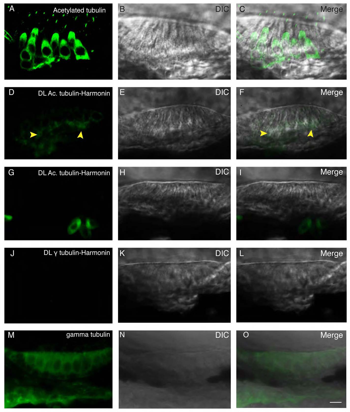
Fig. S9 Proximity ligation assay indicates close physical proximity at the subcellular level. (A) Immunolabeling of acetylated tubulin. Acetylated tubulin fills up the cytoplasm and labels the kinocilium. (B) DIC channel. (C) Merge of panels A and B. (D) Proximity ligation assay of acetylated tubulin and Harmonin results in a restricted signal localized in the basal region of the hair cell, as indicated by the yellow arrowheads. (E) DIC channel. (F) Merge of panels D and E. (G) Proximity ligation assay of acetylated tubulin and Harmonin in ush1c mutant results in the absence of signal. (H) DIC channel. (I) merge of panels G and H. (J) Proximity ligation reaction of gamma tubulin and Harmonin resulted in no signal. (K) DIC channel. (L) Merge of panels J and K. (M) Immunolabeling of gamma tubulin showing a nonspecific signal filling the cytoplasm. (N) DIC channel. O: merge of panels M and N. Scale bar: 5µm.
Image
Figure Caption
Acknowledgments
This image is the copyrighted work of the attributed author or publisher, and
ZFIN has permission only to display this image to its users.
Additional permissions should be obtained from the applicable author or publisher of the image.
Full text @ Dis. Model. Mech.

