Fig. S3
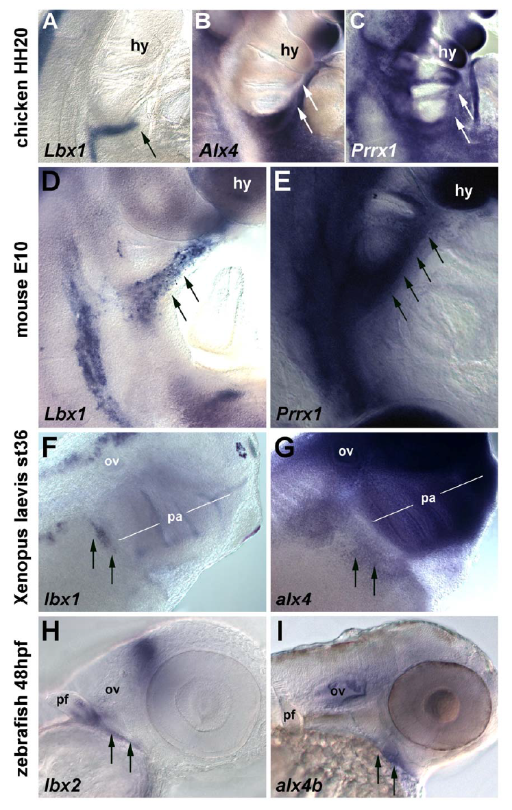
Headtrunk interface of (AC) chicken embryos at HH20, (D,E) mouse embryos at E10.5 pc, (F,G) Xenopus laevis embryos at st36 and (H,I) zebrafish embryos at 48 hpf; lateral views, rostral to the top (AE) or the right (FI); the molecular markers used are indicated. The hypobranchial/hypoglossal muscle precursors (HMP) express Lbx1 (A,D,F, arrows) or in the zebrafish, the paralogous gene lbx2 (H, arrows). The paired type homeobox genes Alx4 and Prrx1 are expressed in the lateral mesoderm, with expression domains anticipating the path of the HMP (B,C,E,G,I arrows).
Abbreviations: hy, hyoid arch; ov, otic vesicle; pa, pharyngeal arches; pf, pectoral fin.
Chicken, mouse and Xenopus represent the sarcopterygian and zebrafish the actinopterygian class of osteichthyans (?bony? jawed vertebrates). The conserved expression patterns shown here suggest a shared developmental mechanism that was present deep in osteichthyan evolution.
In chicken, mouse and Xenopus, Lbx1 labelled the emigrating HMP on their circumpharyngeal path (Suppl. Fig.3A,D,F; arrows). In the zebrafish, while the closely related lbx1a, 1b and 2 genes marked migratory muscle precursors for the pectoral fins (Neyt et al., 2000; Ochi and Westerfield, 2009; Thisse et al., 2004; Wotton et al., 2008) and Wotton and Dietrich, unpublished observations), lbx2 was expressed in HMP (Suppl. Fig.3H, arrows). In chicken, mouse and zebrafish, Alx4 (zebrafish: alx4b) labelled the lateral mesoderm surrounding the most caudal pharyngeal arch and the hypopharyngeal region (Suppl. Fig.3B,G,I; arrows), in the frog accompanied by expression in neural crest cells filling the arches (Suppl. Fig.3G, pa). In the mouse, circumpharyngeal expression of Alx4 was low, but in both chicken and mouse, Prrx1 showed the same, rostrally expanding expression along the floor of the pharynx (Suppl. Fig.3C,E; arrows). Notably, in all species, the lateral mesoderm markers spread into hypopharyngeal areas before the HMP.
Reprinted from Developmental Biology, 390, Lours-Calet, C., Alvares, L.E., El-Hanfy, A.S., Gandesha, S., Walters, E.H., Sobreira, D.R., Wotton, K.R., Jorge, E.C., Lawson, J.A., Kelsey Lewis, A., Tada, M., Sharpe, C., Kardon, G., Dietrich, S., Evolutionarily conserved morphogenetic movements at the vertebrate head-trunk interface coordinate the transport and assembly of hypopharyngeal structures, 231-46, Copyright (2014) with permission from Elsevier. Full text @ Dev. Biol.

