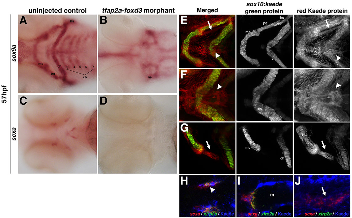
Fig. 4 Zebrafish craniofacial tendon populations are derived from the neural crest. Morpholino-mediated knockdown of tfap2a and foxd3 results in (A,B) complete loss of sox9a-positive pharyngeal cartilage (98%, n=58) and (C,D) scxa-positive craniofacial tendon progenitors (96%, n=46) at 57hpf compared with controls. (E-G) 72hpf photoconverted sox10:kaede embryos express sox10:kaede green protein in the pharyngeal cartilage (E-G and middle panel), and the red Kaede protein from the 22hpf photoconversion is found in the two major populations of craniofacial tendon progenitors (E, arrow and arrowhead; F,G, right panel). (H-J) Colocalization of scxa and Kaede protein in photoconverted sox10:kaede embryos at 72hpf is observed in the sternohyoideus connection point (H,I; sternohyoideus muscle is labeled m) and in the ligament (J). A subset of the scxa-positive and Kaede-positive cells also co-expresses xirp2a transcripts. Arrows in E,G,J mark ligament medial to palatoquadrate; arrowheads in E,F,H mark tendon connecting the sternohyoideus to the ceratohyals. cb: ceratobranchials; ch, ceratohyal; hs, hyosymplectic; m, muscle; mc, Meckel′s cartilage; ne, neurocranium; pq, palatoquadrate.
Image
Figure Caption
Figure Data
Acknowledgments
This image is the copyrighted work of the attributed author or publisher, and
ZFIN has permission only to display this image to its users.
Additional permissions should be obtained from the applicable author or publisher of the image.
Full text @ Development

