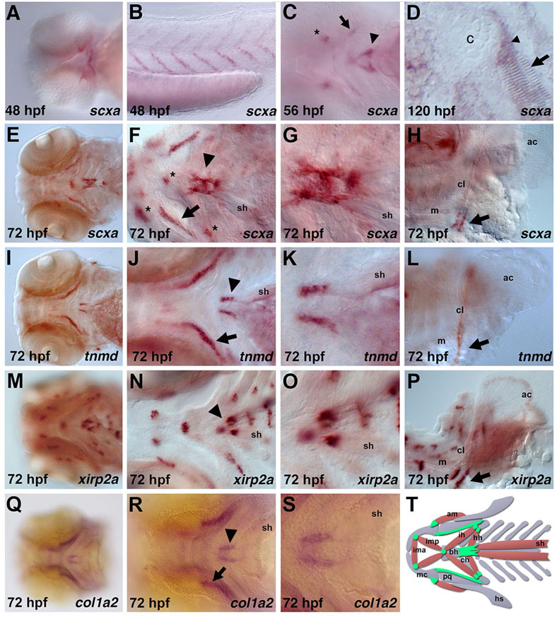
Fig. 1 Expression of tendon markers during zebrafish development. At 48hpf, scxa is expressed in the (A) pharyngeal arches and (B) myosepta. (C) At 56hpf, scxa is expressed in the craniofacial region (asterisk, arrow, arrowhead). (D) Section in situ hybridization of scxa expression (arrowhead) between cartilage (c) and muscle (arrow) at 120hpf. (E-S) Expression of scxa (E-H), tnmd (I-L), xirp2a (M-P) and col1a2 (Q-S) at 72hpf. All four genes are expressed at the attachment point of the sternohyoideus muscles to the ceratohyal and basihyal cartilages (F,J,N,R, arrowhead; magnified in G,K,O,S). scxa, tnmd and col1a2 are robustly expressed in two stripes ventromedial to the palatoquadrate (F,J,R, arrow). scxa and xirp2a are expressed at the adductor mandibulae, intermandibularis and hyohyoideus muscle attachment points (F, asterisks; N). scxa, tnmd and xirp2a are also expressed at the base of the cleithrum (H,L,P, arrow). (T) Schematic ventral view of zebrafish craniofacial muscle (red), cartilage (gray) and tendon/ligament (green) populations at 72hpf. Only subsets of the muscle groups are depicted. All are ventral views of flat-mounted embryos except in B,H,L,P, which are lateral views, and in D, which is a coronal view. ac, actinotrichia; am, adductor mandibulae; bh, basihyal; c, cartilage; cl, cleithrum; ch, ceratohyal; hh, hyohyoideus; ih, interhyoideus; ima, intermandibularis anterior; imp, intermandibularis posterior; m, muscle; mc, Meckel′s cartilage; pq, palatoquadrate; sh, sternohyoideus.
Image
Figure Caption
Figure Data
Acknowledgments
This image is the copyrighted work of the attributed author or publisher, and
ZFIN has permission only to display this image to its users.
Additional permissions should be obtained from the applicable author or publisher of the image.
Full text @ Development

