Fig. S5
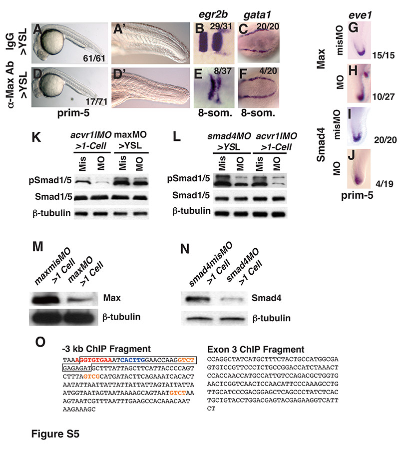
Embryos injected into the YSL with 150pg IgG (A-C), 150pg anti-Max antibody (D-E), 8ng maxmisMO (G, K, M), 8ng maxMO (H, K, M), 4ng smad4misMO (I, L, N) or 4ng smad4MO (J, L, N). (A, A′, D, D′) Image of live embryo at prim-5 (24 hpf). (B, E) egr2b expression at the 8- somite stage. (C, F) gata1 expression at the 8-somite stage. (G, H, I, J) eve1 expression in the tailbud at the prim-5 stage. Arrowhead indicates ectopic tailbud. (K) Western Blot of extracts of embryos at 75% epiboly acvr1lMO>1-cell morphants, maxMO>YSL morphants or controls. In acvr1lMO>1-cell morphants, pSmad1: 25% ± 8.6 S.D. of controls; pSmad5: 21.25% ± 4.7 S.D. of controls; In maxMO>YSL morphants, pSmad1: 56% ± 6.3 S.D. of controls, pSmad5: 70% ± 7.0 S.D. of controls. (L) Western Blot of extracts of embryos at 75% epiboly acvr1lmisMO>1-cell morphants, smad4MO>YSL morphants, or controls. In acvr1lMO>1-cell morphants, pSmad1: 25% ± 8.6 S.D. of controls; pSmad5: 21.25% ± 4.7 S.D. of controls; In smad4MO>YSL morphants, pSmad1: 23% ± 3.5 S.D. of controls, pSmad5: 35% ± 0.7 S.D. of controls. (M) Western blot of maxMO>1-cell morphants or controls. Max protein: 19.7% ± 7.3 S.D. of controls. (N) Western blot of extracts of 8hpf smad4MO>1-cell morphants or controls. Smad4 protein: 20% ± 2.1 S.D. of controls. (O) The sequences of the amplicons assayed in Fig. 7A. Consensus Tbox site is in red; consensus E-box site is blue; potential Smad binding sites are orange. Anterior is to the left in (A-F), to the top in (G, H, I, J). For Western blots, 15μg total protein loaded in each lane.
Reprinted from Developmental Cell, 28(3), Sun, Y., Tseng, W.C., Fan, X., Ball, R., and Dougan, S.T., Extraembryonic signals under the control of MGA, Max, and Smad4 are required for dorsoventral patterning, 322-334, Copyright (2014) with permission from Elsevier. Full text @ Dev. Cell

