
|
Fig. S1
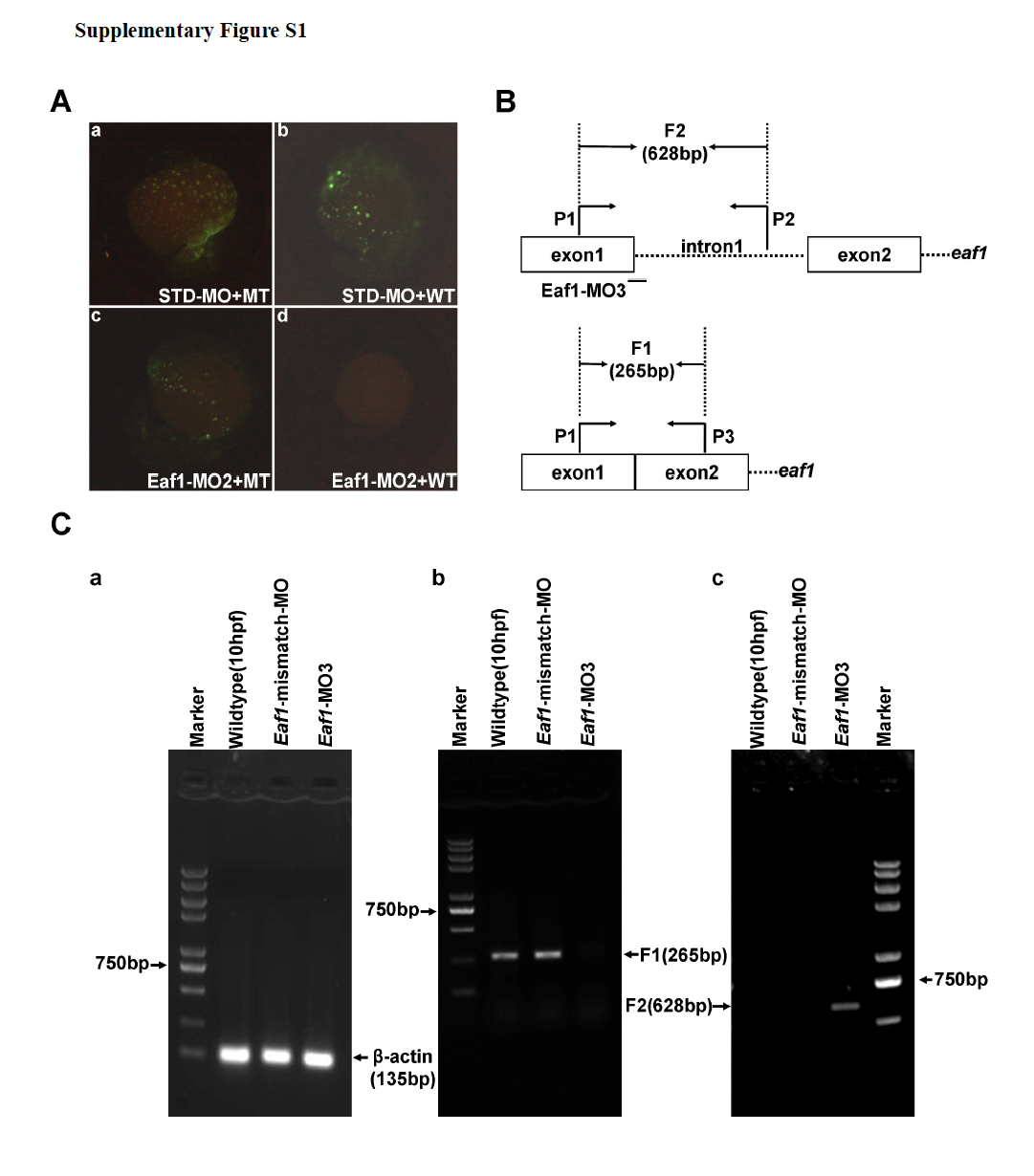
Validation of Eaf1-MO2 and Eaf1-MO3. (A) Embryos co-injected with the standard morpholinoSTD and the MT reporter vector (mutated Eaf1-MO targeted region fused with GFP) or WT reporter vector (wildtype Eaf1-MO targeted region fused with GFP) demonstrated that STD could not block expression of the GFP fusion protein (a and b); embryos co-injected with Eaf1-MO2 and the MT reporter or WT reporter indicated that Eaf1-MO2 could not block expression of MT-GFP fusion protein (c), but could block expression of WT-GFP fusion protein effectively (d). (B) The schematic of eaf1 exon-intron structure, Eaf1-MO3 targeting position and RT-PCR primer locations. (C) (a) A 135-bp β-actin fragment was detected in all embryos by RT-PCR. (b) An expected 265-bp fragment resulted from normal splicing of eaf1 could be detected by RT-PCR in the wildtype embryos and the embryos injected with Eaf1-mismatch-MO (8ng/per embryo), but not in the embryos injected with Eaf1-MO3 (4ng/per embryo). (c) An expected 628-bp fragment resulted from splice-blocking of eaf1 could be detected by RT-PCR in the embryos injected with EAF1-MO3, but not in the wildtype embryos and the embryos injected with Eaf1-mismatch-MO. The embryos were collected at 9-10 hpf.
Reprinted from Developmental Biology, 388(1), Hu, B., Zhang, W., Feng, X., Ji, W., Xie, X., and Xiao, W., Zebrafish eaf1 suppresses foxo3b expression to modulate transcriptional activity of gata1 and spi1 in primitive hematopoiesis, 81-93, Copyright (2014) with permission from Elsevier. Full text @ Dev. Biol.