
Image
|
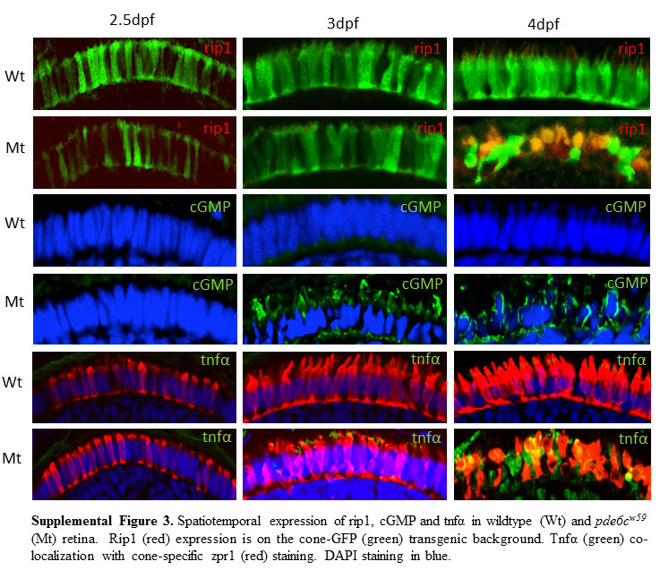
Figure Caption
Fig. S3
Spatiotemporal expression of rip1, cGMP and tnfα in wildtype (Wt) and pde6cw59 (Mt) retina. Rip1 (red) expression is on the cone-GFP (green) transgenic background. Tnfα (green) co-localization with cone-specific zpr1 (red) staining. DAPI staining in blue.
Acknowledgments
This image is the copyrighted work of the attributed author or publisher, and
ZFIN has permission only to display this image to its users.
Additional permissions should be obtained from the applicable author or publisher of the image.
Full text @ Cell Death Differ.