
Fig. 4
- ID
- ZDB-IMAGE-140416-37
- Genes
- Antibodies
- Source
- Figures for Viringipurampeer et al., 2014
|
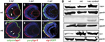
Fig. 4
Expression patterns of caspase-independent cell death markers. (a–c) Wild-type (wt) retinal images. (d–f) Mutant pde6c-/- (mt) retinal images. (a and d) Double labelling of calpain in green and zpr1 cone-specific marker in red. A few cells express calpain in the inner retina (white arrows). Scale bar=20μm. (b ande) Double labelling of parp in green and zpr1 in red. (c and f) rip1 co-labelling in red on GFP-expressing cone transgenic background. (g) Western blot of calpain (capn), parp and rip1 at 7d.p.f. Positive (+ve) controls: for parp and capn, mouse E14.5 eye tissue; for rip1, HEK293 cell line. Gapdh used to normalize gel loading