Image
Figure Caption
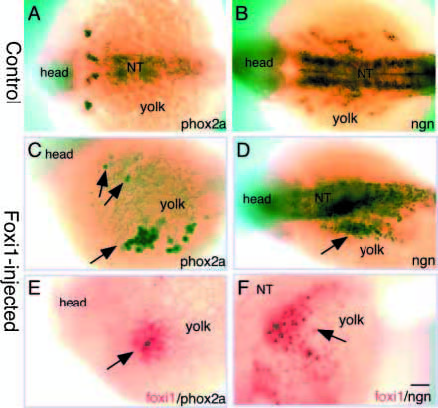
Fig. 7 Ectopic expression of foxi1. Anterior is to the left. (A,B) Dorsal views of control embryos labeled with phox2a and ngn antisense RNA probes, respectively. (C-F) 24 hpf foxi1-injected embryos, labeled with (C) phox2a, showing ectopic phox2a+ cells on the yolk surface (arrows); (D) ngn, showing ectopic ngn+ cells on the yolk surface (arrow); (E) foxi1 (red) and phox2a (purple), showing that phox2a+ cells do express ectopic foxi1; (F) foxi1 (red) and ngn (purple), showing that ngn+ cells do express ectopic foxi1. Scale bar, 100 μm.
Acknowledgments
This image is the copyrighted work of the attributed author or publisher, and
ZFIN has permission only to display this image to its users.
Additional permissions should be obtained from the applicable author or publisher of the image.
Full text @ Development

