Fig. 6
NLRC3-like Negatively Regulates Inflammatory Signaling through Macrophages and May Interact with the Inflammasome Component ASC and Other Pyrin and NACHT-Containing Proteins
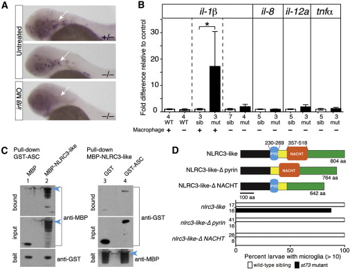
(A) Macrophage mfap4 expression shows complete macrophage ablation by irf8 MO injection at 2 dpf (arrow). Injected wild-type embryos show complete loss of neutral red-positive microglia (n = 29/29; data not shown).
(B) Graph shows relative il-1β mRNA levels, with pairwise comparisons indicated by the dotted lines. Depletion of macrophages in nlrc3-like mutants (mut) reduces il-1β and other proinflammatory cytokine levels similar to wild-type (WT) levels. sib, siblings.
(C) SDS-PAGE analyses of reciprocal pull-down assays show binding of zebrafish ASC with NLRC3-like (lanes 2 and 4) and minimal binding to tag-alone controls (lanes 1 and 3). MBP-tagged full-length NLRC3-like runs near 160 kDa (blue arrowhead) with several smaller processed forms; GST-ASC at 50 kDa; MBP alone from pMAL-c2X vector at 50 kDa; and GST alone at 30 kDa. Blot shows 0.4% of the input prey protein per lane.
(D) Top view is a schematic of the wild-type NLRC3-like protein and deletion versions: NLRC3-like- δpyrin with deletion at amino acids (aa) 230?269 and NLRC3-like-δNACHT with deletion at aa 357?518. At the bottom is a bar graph showing rescue of st73 mutants (87.5%, n = 16) by injection of mRNA encoding wild-type NLRC3-like, but none using the mutant constructs. Numbers at left edge of bar graphs represent n, number of embryos analyzed.
Error bars show SEM. p < 0.05.

