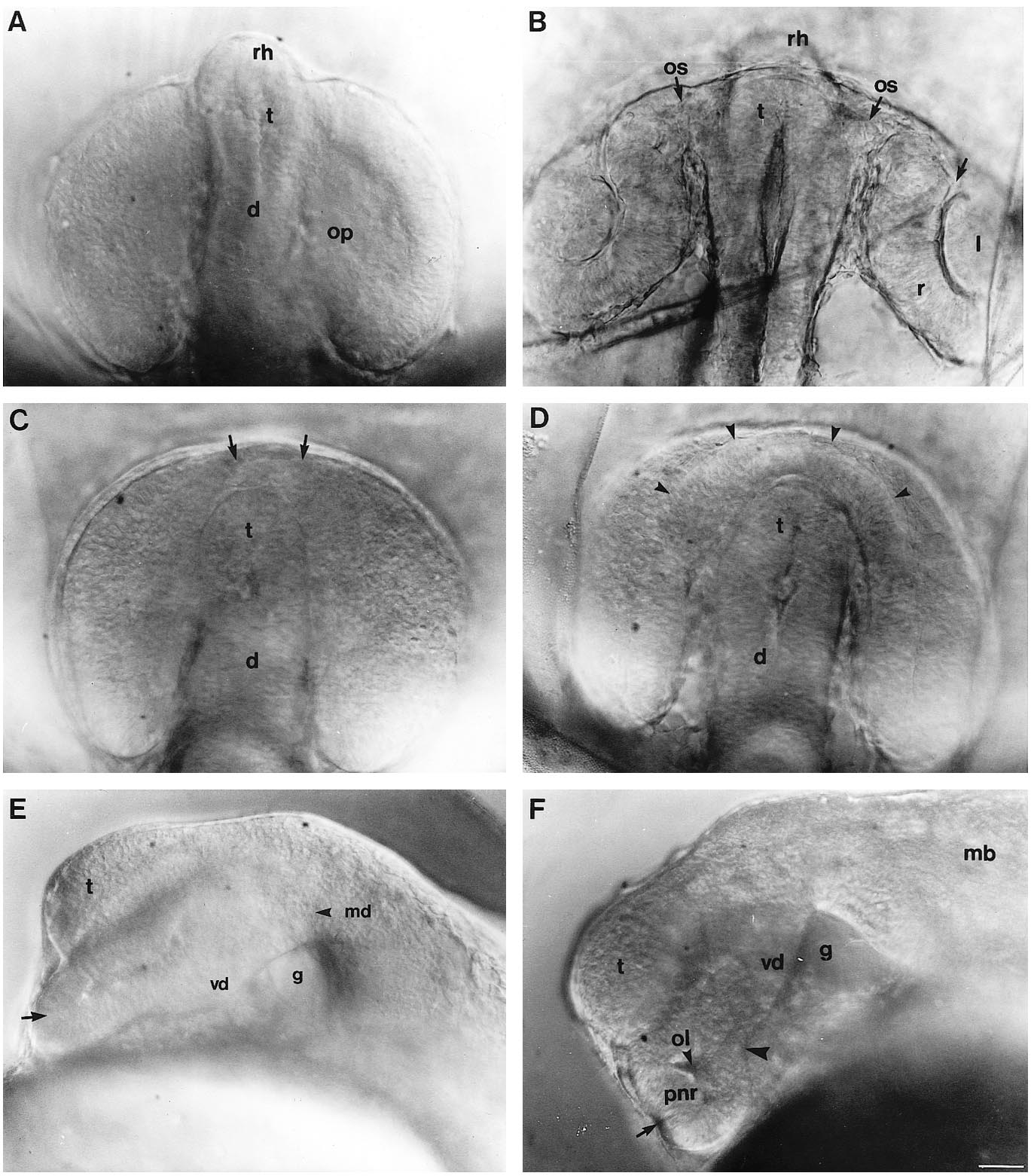
Fig. 1 Development of the eye in living wild-type embryos and embryos homozygous for the cyclops mutation. (A,B) Wildtype embryos, (C-F) cyclops embryos. (A) 13s, dorsal view. (B) 24 hour, dorsal view. The arrow indicates the rostral limit of optic cup invagination. The position of the telencephalon is indicated although it is above the plane of focus. (C) 13s, dorsal view. The arrows indicate the bridge of fused retina around the anterior pole of the neural keel. The anterior hypothalamus (see A and B) is missing from mutant embryos. (D) 20-22s, dorsal view. The arrowheads indicate minor invagination in the bridge of fused neural retina. (E) 16s, lateral view focused on the midline. The arrow indicates the area of prospective retinal fusion. A large gap is present beneath the diencephalon. (F) 20-22s, lateral view focused on the midline. The arrow indicates a small amount of invagination in the bridge of prospective retinal tissue. The arrowhead indicates the caudal limit of retinal fusion. Abbreviations: d, diencephalon; g, gap in mid-diencephalic neuroepithelium; l, lens; mb, midbrain; md, mid-diencephalon; ol, optic lumina; op, optic primordia; os, optic stalks; pnr, presumptive neural retina; r, retina; rh, rostral hypothalamus; t, telencephalon; vd, ventral diencephalon. Scale bar, 25 μm.
Image
Figure Caption
Figure Data
Acknowledgments
This image is the copyrighted work of the attributed author or publisher, and
ZFIN has permission only to display this image to its users.
Additional permissions should be obtained from the applicable author or publisher of the image.
Full text @ Development

