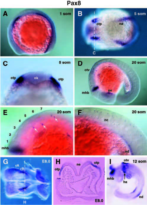
Fig. 6 Identification of novel expression domains of the vertebrate Pax8 gene. (A) Early Pax8 expression in the intermediate mesoderm (im) and in the anlage of the otic placode (otp). (B) Dorsal view of a 5-somite embryo with lateral expression of Pax8 in the otic placode and pronephric anlage (na). (C) Optical cross-section of the 5-somite embryo at the level of the otic placode (as indicated in B), which localizes the Pax8 staining in the ectoderm adjacent to the neural keel (nk). Lateral view (D) and higher magnifications (E,F) of a 20- somite embryo. Pax8 expression is no longer observed in the otic vesicle (otv), while it is maintained in the pronephros (out of focal plane) and nephric duct (nd). Arrows in E indicate individual Pax8- expressing neurons in the hindbrain, and numbers refer to individual rhombomeres. (G) Dorsal view of an 8.0-day mouse embryo (before somitogenesis) hybridized with a mouse Pax8 RNA probe. (H) Cross-section through the same embryo at the level indicated in G. Pax8 staining is detected lateral to the neuroepithelium (ne) of rhombomere B (rB) in the ectodermal layer corresponding to the otic placode. (I) Mouse Pax8 expression at the 12-somite stage. ha, hyoid arch; nc, notocord; ov, optic vesicle.
Image
Figure Caption
Figure Data
Acknowledgments
This image is the copyrighted work of the attributed author or publisher, and
ZFIN has permission only to display this image to its users.
Additional permissions should be obtained from the applicable author or publisher of the image.
Full text @ Development

