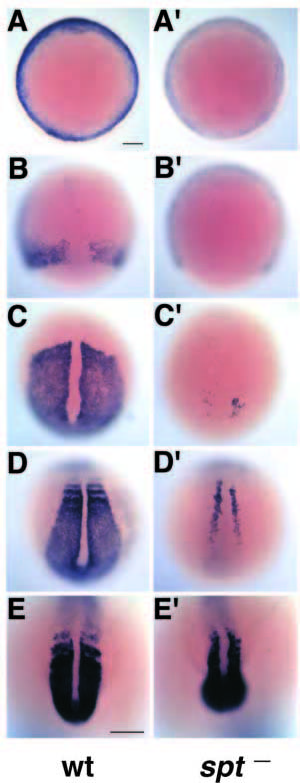
Fig. 3 papc expression is defective in spt mutant embryos. (A-E) Wild-type embryos. (A′-E′) sptb104 homozygous mutant embryos. (A,A′) Shield stage showing greatly reduced papc in spt- embryos at the onset of gastrulation. (B,B′) 80% epiboly showing lack of papc expression in the marginal zone of the spt mutant. (C,C′) Bud (10 hour) stage, (D,D′) 4-6 somite stage; expression is detected only in a few adaxial-like cells in spt mutants. (E,E′) 18 somite stage; the expression of papc recovers in tail somites, in the segmental plate and in the tailbud of spt mutants. A-D and A′-D′ are dorsal views, E and E′ are posterior views. Bars in A (also applies to B-D) and E, 100 μm.
Image
Figure Caption
Figure Data
Acknowledgments
This image is the copyrighted work of the attributed author or publisher, and
ZFIN has permission only to display this image to its users.
Additional permissions should be obtained from the applicable author or publisher of the image.
Full text @ Development

