
Image
|
Figure Caption
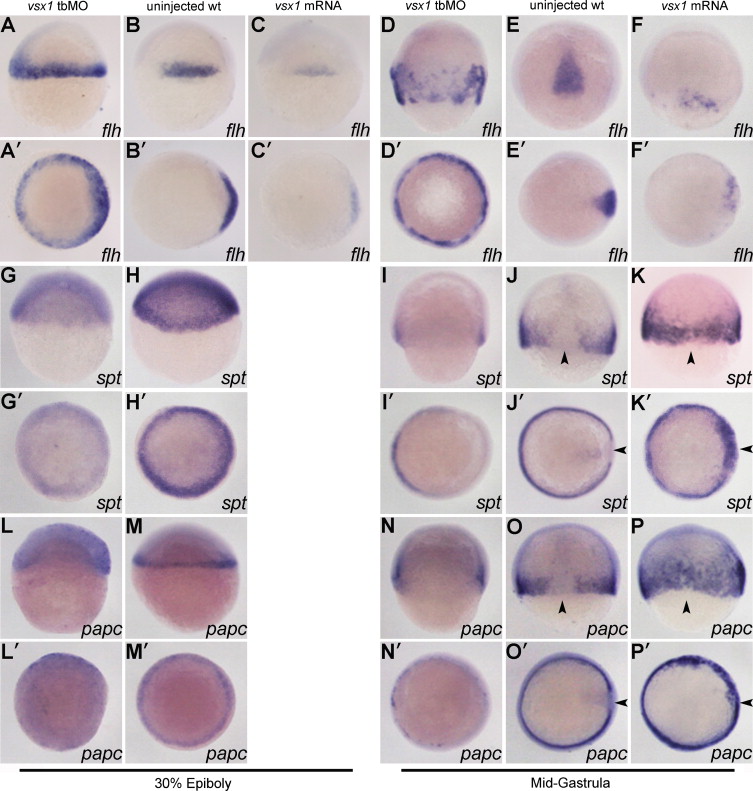
Fig. 5
Vsx1 represses flh to preserve spt and papc expression in the ventolateral margin. The injected reagents are indicated at the top of each column. Riboprobes are indicated at the bottom of each figure. (A?P) Dorsal view of embryos with animal pole towards the top. (A′?P′) Animal pole view of the embryos with dorsal towards the right. Arrow heads indicate that the paraxial mesoderm marker is detected in the presumptive axial mesoderm region in vsx1 overexpression embryos.
Figure Data
Acknowledgments
This image is the copyrighted work of the attributed author or publisher, and
ZFIN has permission only to display this image to its users.
Additional permissions should be obtained from the applicable author or publisher of the image.
Reprinted from Developmental Biology, 386(1), He, Y., Xu, X., Zhao, S., Ma, S., Sun, L., Liu, Z., and Luo, C., Maternal control of axial-paraxial mesoderm patterning via direct transcriptional repression in zebrafish, 96-110, Copyright (2014) with permission from Elsevier. Full text @ Dev. Biol.