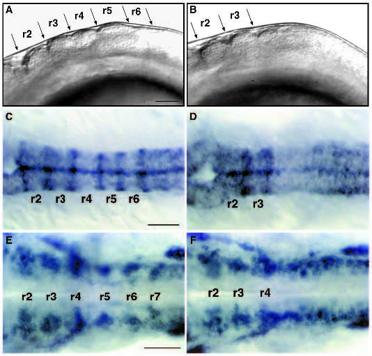
Fig. 2 val- embryos lack segment boundaries and segmental patterns of neuronal differentiation posterior to rhombomere 4. (A,B) Lateral view of live 18 h wild-type (A) and val- (B) embryos. Anterior is to the left. In val- embryos, there are no visible rhombomere boundaries posterior to the r3-r4 boundary. (C,D) Dorsal views of RNA in situ hybridizations of wild-type (C) and val- (D) embryos at 18 h showing expression of mariposa in the rhombomere boundaries. No expression is observed posterior to the r3-r4 boundary in val- embryos. (E,F) Dorsal views of RNA in situ hybridizations of wildtype (E) and val- (F) embryos at 24 h showing expression of gap43 in clusters of early differentiating neurons laterally in each rhombomere. In val- embryos, this segmental pattern of gap43 staining is lost posterior to r4. This disrupted pattern of neuronal differentiation is also observed in val- embryos stained with the HNK-1 antibody (data not shown; Metcalfe et al., 1990; Trevarrow et al., 1990). Scale bars, 50 μm.
Image
Figure Caption
Figure Data
Acknowledgments
This image is the copyrighted work of the attributed author or publisher, and
ZFIN has permission only to display this image to its users.
Additional permissions should be obtained from the applicable author or publisher of the image.
Full text @ Development

