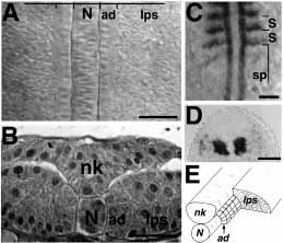
Fig. 1 Adaxial and lateral presomitic cells can be distinguished in the segmental plate. (A) Dorsal view of the segmental plate of an approximately 13 h (8 somites have formed) live embryo. The cuboidal adaxial cells (ad) on either side of the notochord (N) can be distinguished from the irregularly shaped lateral presomitic cells (lps). (B) Transverse semithin section of the segmental plate of a 12 h (6 somites) embryo. The adaxial cells (ad) are large cells adjacent to the notochord (N). Lateral presomitic cells (lps) are smaller, more irregularly shaped and not contacting the notochord. nk, neural keel. (C) Dorsal view of a 13 h (8 somites) embryo labeled by wholemount RNA in situ hybridization for myoD. In the segmental plate (sp), only adaxial cells abundantly express myoD; as somites (S) form, other somitic cells also express myoD. Horizontal lines mark somite borders. (D) Transverse section through the most rostral portion of the segmental plate of an approximately 15 h (12 somites) embryo labeled by whole-mount in situ hybridization for myoD. Adaxial cells express very abundant levels of myoD. Some of the lateral presomitic cells are beginning to express low levels of myoD. (E) Schematic drawing of the segmental plate. The adaxial cells (ad) are arranged as a sheet between the notochord (N) and the lateral presomitic cells (lps); the neural keel (nk) is also shown. Approximately 20 adaxial cells contribute to each somite. In dorsal views, anterior is to the top of the figure; in transverse sections, dorsal is up. Scale bars: A,B, 25 μm; C, D, 50 μm.
Image
Figure Caption
Figure Data
Acknowledgments
This image is the copyrighted work of the attributed author or publisher, and
ZFIN has permission only to display this image to its users.
Additional permissions should be obtained from the applicable author or publisher of the image.
Full text @ Development

