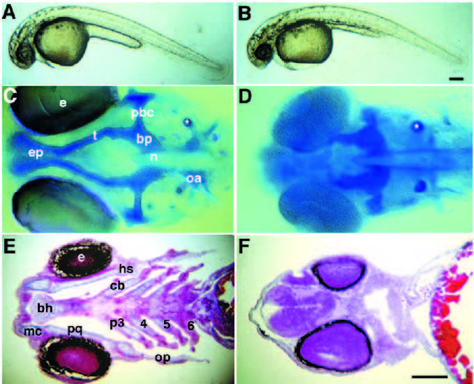
Fig. 1 Embryos homozygous for the chn mutation lack much of the ventral head skeleton and striated musculature. (A,C,E) Wild type; (B,D,F) mutants. (A,B) 30 hour, lateral view. Living wild-type and chnb146 embryos at the prim-15 stage. chn mutants have smaller eyes, a pointed nose and slightly irregular yolks. (C,D) 72 hour, ventral view. By late hatching, much of head skeletal development is blocked. Whole-mounted embryos labelled with Alcian blue. The basilar plate (bp) flanking the anterior notochord is often shorter and thicker than in the wild type. Otoliths within the otic capsule also stain (asterisk). (E,F) 96 hour. Horizontal sections (7 mm) through the pharyngeal region, stained with Mallory’s triple stain for cartilage and muscle (Pantin, 1960). In wild-type embryos, jaw extension moves the mouth opening anterior to the eyes and seven pharyngeal segments differentiate. A mouth opening forms but remains in its original posterior location in chn mutants, surrounded by undifferentiated mesenchyme. All pharyngeal cartilages (blue) as well as striated muscles (red) are absent in chn. Abbreviations: bh, basihyal; cb, ceratobranchials; e, eyes; ep, ethmoid plate; hs, hyosymplectic; mc, Meckel’s cartilage; oa, occipital arch; op, operculum; pbc, posterior basicapsular commissure; p3-6, pharyngeal arches 3-6; pq, palatoquadrate; t, trabeculae cranii. Scale bars, 200 μm.
Image
Figure Caption
Figure Data
Acknowledgments
This image is the copyrighted work of the attributed author or publisher, and
ZFIN has permission only to display this image to its users.
Additional permissions should be obtained from the applicable author or publisher of the image.
Full text @ Development

