
|
Fig. 1
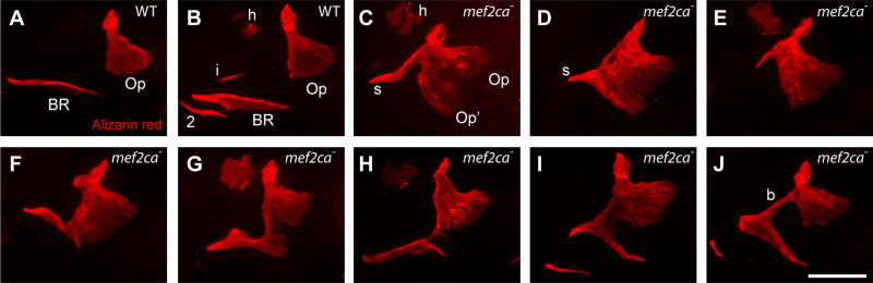
The shapes of the opercle (Op) ? branchiostegal ray 3 (BR) bones are outstandingly diverse in mef2ca mutants. Confocal projections (dorsal up, anterior to the left), bones vitally stained with Alizarin Red and live-imaged at 6 dpf. (A and B) wild type (WT), with (B) evidently the more advanced (h: hyomandibula, i: interopercle rudiment, and 2: a second BR). (C?J) mef2ca mutants. The expanded mutant bone in (C) appears to a mirror-image duplicated Op (Op2), and a strut (s) of bone may, or may not, include a BR rudiment. Examples of mutants in (F?J) include the BR bridged to the OP (b, panel J). About 2/3 of the mutants in the single pair family used for this and Fig. 2 include either a strut or bridge as part of the ectopic bone formation. Scale bar 50 μm.
Reprinted from Developmental Biology, 385(2), Delaurier, A., Huycke, T.R., Nichols, J.T., Swartz, M.E., Larsen, A., Walker, C., Dowd, J., Pan, L., Moens, C.B., and Kimmel, C.B., Role of mef2ca in developmental buffering of the zebrafish larval hyoid dermal skeleton, 189-99, Copyright (2014) with permission from Elsevier. Full text @ Dev. Biol.