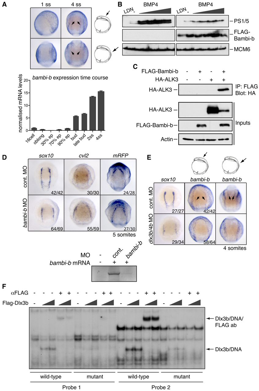
Fig. 6 dlx3b induces the expression of bambi-b in the PPE to suppress localised BMP activity and allow tissue specification at the NPB. (A) Temporal expression analysis of bambi-b by ISH (top) and qRT-PCR (bottom). Two orientations (indicated by arrows on the drawings) are shown for the same embryo at each stage (top row in anterior view with anterior to the top). Arrowheads indicate the stripes of bambi-b expression at the NPB. Error bars indicate s.d. of triplicates. (B) HEK293T cells transfected with control plasmid or pCS2+-FLAG-Bambi-b were treated with different concentrations of BMP4 (2, 4, 10 and 20 ng/ml) for 1 hour, or with the BMP receptor inhibitor LDN-193189 for 3 hours. Whole cell extracts were western blotted using the indicated antibodies. PS1/5, PSmad1/5. (C) HEK293T cells were transfected with control plasmid or pCS2+-FLAG-Bambi-b or HA-ALK3. FLAG-Bambi-b was immunoprecipitated from whole cell extract and the immunoprecipitates were western blotted for HA. The inputs are also shown. Actin provided a loading control. (D) (Top) ISH for sox10, cvl2 and mRFP on bambi-b morphants as compared with controls. Embryos were injected with 10 ng bambi-b or control MO and fixed at the 5-somite stage. Dorsal anterior view, anterior to the top. (Bottom) Synthetic mRNA corresponding to native bambi-b was translated in reticulocyte lysate in the presence of 0.2 mM bambi-b MO and [35S]methionine. Translation products were separated by SDS-PAGE and visualised by autoradiography. (E) ISH for sox10 and bambi-b on dlx3b/4b morphants compared with control MO-injected embryos fixed at the 4-somite stage. Views and arrows/arrowheads are as in A. (F) FLAG-Dlx3b expressed in HEK293T cells was assessed for binding to DNA using a bandshift assay. The wild-type probes correspond to the two putative Dlx3b binding sites in the bambi-b gene and the mutant probes contain point mutations in the binding sites. The Dlx3b-DNA complexes are indicated, as are the supershifted complexes that additionally contain the anti-FLAG antibody. ep, epiboly; ss, somitogenesis stage.
Image
Figure Caption
Figure Data
Acknowledgments
This image is the copyrighted work of the attributed author or publisher, and
ZFIN has permission only to display this image to its users.
Additional permissions should be obtained from the applicable author or publisher of the image.
Full text @ Development

