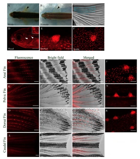
Fig. S1 Distribution and morphology of BTs in male zebrafish. (A) Dorsal view of a male zebrafish with pectoral fin BTs (black arrowhead). (B) Dorsal view of a female zebrafish with no BTs and translucent fins (black arrowhead). (C) Pectoral fin of a male longfin mutant with long BT clusters (outlined in blue). (D) Grouped BTs in a line/semi circle (white box) and isolated BTs (white arrowheads) on the side of a male zebrafish head. (E) BT on a barbel. (F) A BT on a scale pulled from the body surface. (G-J) BTs distributed along the anal fin rays. (K-N) BTs along the pelvic fin rays. (O-R) BTs along the dorsal fin rays. (S-U) No BTs are observed on the caudal fin. Fluorescence Tg(KR21) images (red), Merged (Tg(KR21) + Brightfield) images. Scale bar for C = 200μm; D = 400μm; E = 12.5Ám; J, N, R, F = 25μm; G-I, K-M, O-Q, S-U = 200μm.
Image
Figure Caption
Acknowledgments
This image is the copyrighted work of the attributed author or publisher, and
ZFIN has permission only to display this image to its users.
Additional permissions should be obtained from the applicable author or publisher of the image.
Full text @ Development

