
|
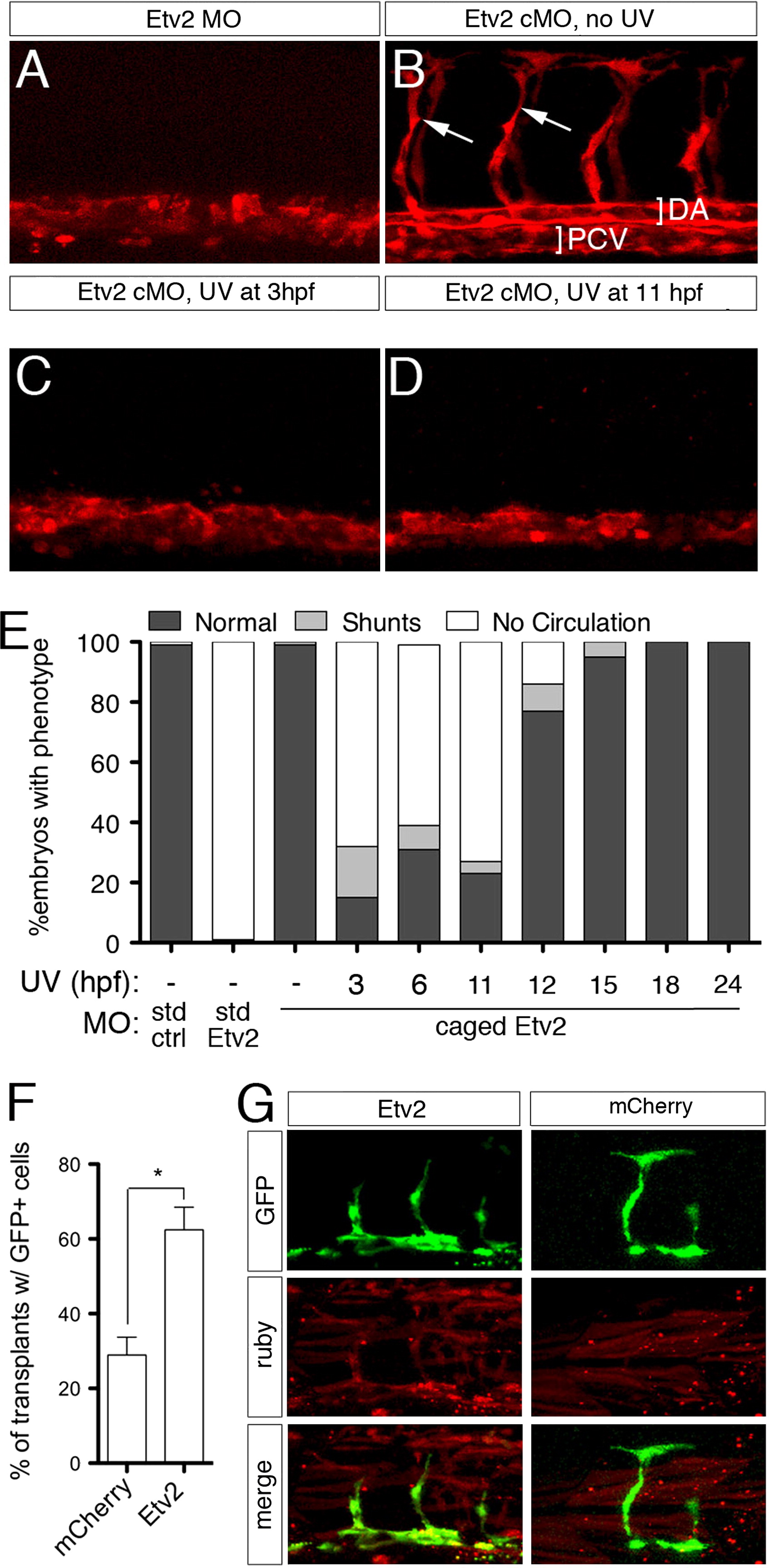
Fig. 2 Etv2 is required only during early stages of vascular development. (A?D) Confocal images of trunk blood vessels in Tg(fli1a.ep:DsRedex)um13 embryos at 30 hpf. Lateral views, dorsal is up, anterior to the left. Embryos injected with (A) 5 ng standard Etv2 Morpholino (MO) or (B) 2 ng Etv2 caged MO (cMO), but not illuminated with UV light. ISVs (arrows), dorsal aorta (DA; bracket) and posterior cardinal vein (PCV; bracket) are indicated. (C, D) Embryos injected with Etv2 cMO exposed to UV light at (C) 3 hpf or (D) 11 hpf. (E) Penetrance of indicated circulatory defects in embryos at 48 hpf following injection with MO and UV exposure as indicated. (F) Percentage of mosaic miniRuby-positive host embryos showing successful transplantation of Tg(fli1:EGFP)y1 donor cells. Donor embryos were injected with 100 pg of mcherry or etv2 mRNA. *p<0.05. (G) Representative confocal images of wild type hosts with contribution to both vascular (green) and non-vascular (red) tissue; donor embryos were injected with mRNA encoding either Etv2 or mCherry.
Reprinted from Developmental Biology, 384(1), Moore, J.C., Sheppard, S., Shestopalov, I.A., Chen, J.K., and Lawson, N., Post-transcriptional mechanisms contribute to Etv2 repression during vascular development, 128-40, Copyright (2013) with permission from Elsevier. Full text @ Dev. Biol.