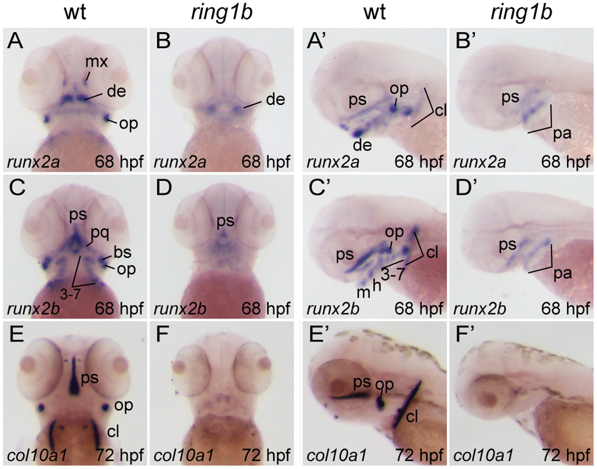
Fig. 6 Loss of endochondral and dermal ossification in ring1b mutants.
Ventral (A?D, E, F) and lateral (A′?D′, E′,) views of in situ hybridizations with riboprobes against the indicated genes in WT and ring1b mutants at 68?72 hpf. In WT embryos, runx2a and runx2b are expressed in hypertrophic pharyngeal arch-derived chondrocytes, as well as in the dermal ossification centers of the operculum, parasphenoid and cleithrum. Weak expression is detected in pharyngeal arches and the parasphenoid of ring1b mutants (B, B′, D, D′). At 72 hpf, col10a1 is expressed in developing dermal bones in WT embryos, but not in ring1b mutants (E?F). cl: cleithrum; de: dentary; h: hyoid; m: mandibular; mx: maxilla; pa: pharyngeal arches; pq: palatoquadrate; ps: parasphenoid, op: operculum cl: the cleithrum. Numbers indicate the respective pharyngeal arches.

