
|
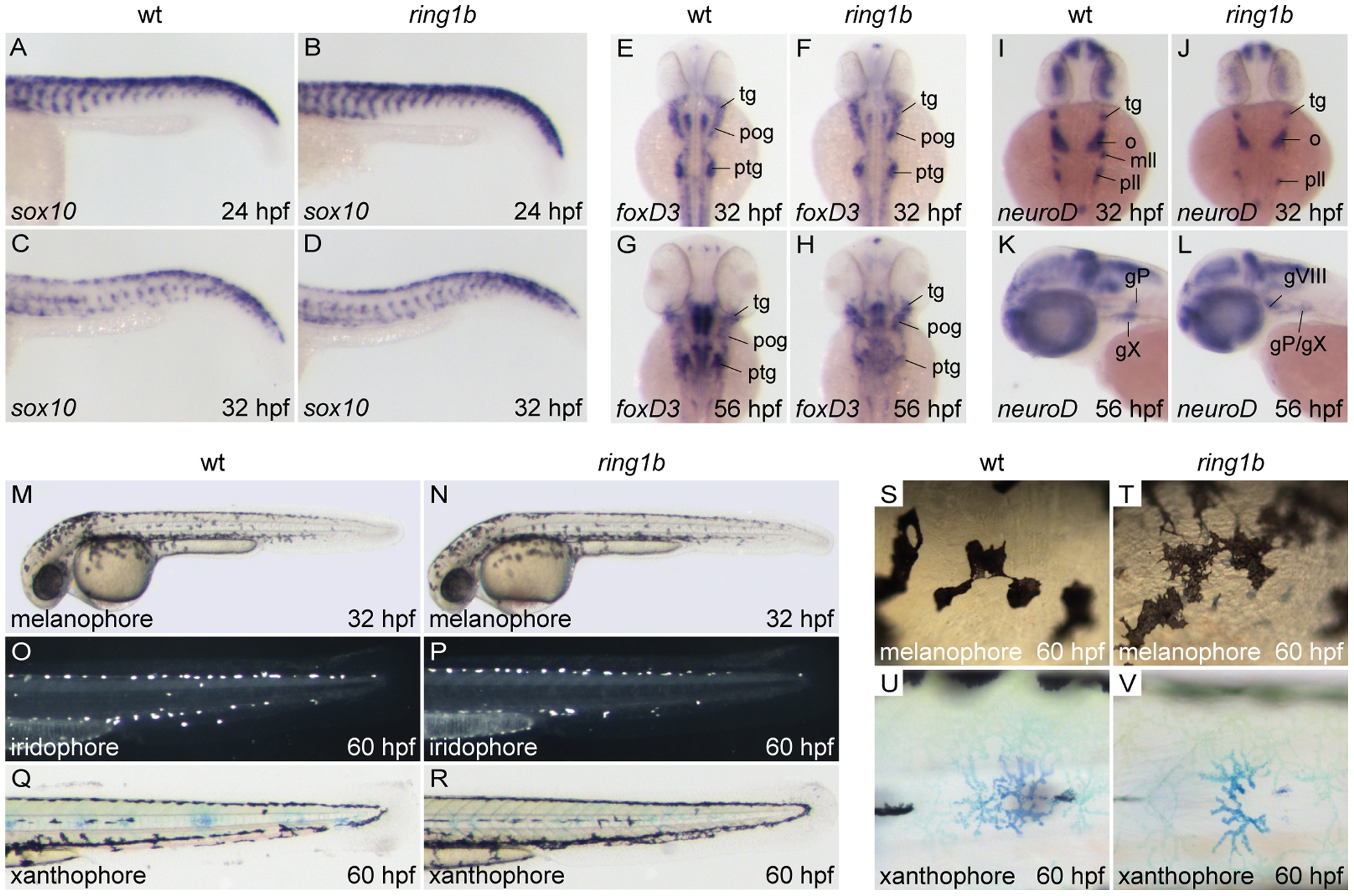
Fig. 3 Migration and initial differentiation of trunk neural crest is largely unaffected in ring1b mutants.
Sox10 staining of WT and ring1b mutants at 24 and 32 hpf shows that sox10 expressing NC cells have migrated timely and correctly to their ventral positions (A–D). Expression of foxD3 in cranial ganglia-associated glia is comparable to WT siblings in ring1b mutants at 32 hpf (E, F), but reduced in the postotic ganglia of ring1b mutants at 56 hpf (G, H). NeuroD expression in cranial ganglia precursors is reduced in ring1b mutants at 32 hpf (I, J), but remains detectable in the hindbrain region of ring1b mutants at 56 hpf (L). Lateral view of 32 hpf embryos shows normal distribution of melanocytes in ring1b mutants at 32 hpf (M, N). At 60 hpf, ring1b melanophores remain more stellate than WT melanophores, which have started to round up (S, T). Iridophores are present in near normal numbers in ring1b mutants at 60 hpf (O, P), whereas ring1b xanthophores are smaller and less stellate (Q, R, U, V). Abbreviations: gX: vagal ganglia; mll: medial lateral line ganglia; o/gVIII: octaval/statoacustic ganglia; pll/gP: posteriolateral line ganglia; pog: preotic ganglia; ptg: postotic ganglia; tg: trigeminal ganglia.