
Fig. 1
|
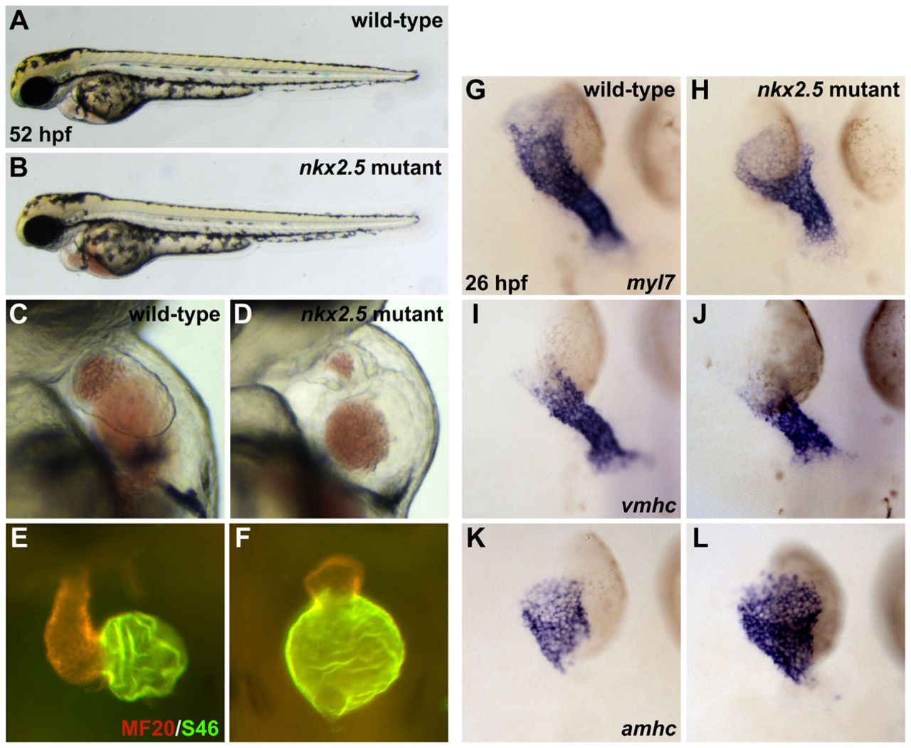
Fig. 1 Mutation of nkx2.5 disrupts cardiac morphogenesis. (A,B) Lateral views of live zebrafish embryos, anterior to the left, at 52 hpf. Other than cardiac defects and pericardial edema, the nkx2.5 mutant embryo (B) appears morphologically normal. (C,D) Lateral views of live embryos, anterior to the top, at 52 hpf. In contrast to the wild-type heart (C), the nkx2.5 mutant heart (D) is unlooped and has striking defects in both ventricular and atrial morphology. (E,F) MF20/S46 immunofluorescence distinguishes ventricular myocardium (red) from atrial myocardium (yellow). Frontal views, anterior to the top, at 52 hpf. In comparison to the wild-type heart (E), the nkx2.5 mutant heart (F) has a diminutive ventricle and an enlarged atrium. (G-L) In situ hybridization depicts expression of myl7 (G,H), vmhc (I,J) and amhc (K,L) in wild-type (G,I,K) and nkx2.5 mutant (H,J,L) embryos. Dorsal views, anterior to the top, at 26 hpf. (G,H) nkx2.5 mutant embryos exhibit subtle defects in heart tube extension, including a broad inflow region and a compact outflow region. In nkx2.5 mutants, the ventricular portion of the heart tube is abnormally short and wide (I,J) and the atrial portion of the heart tube has a splayed appearance (K,L).