
|
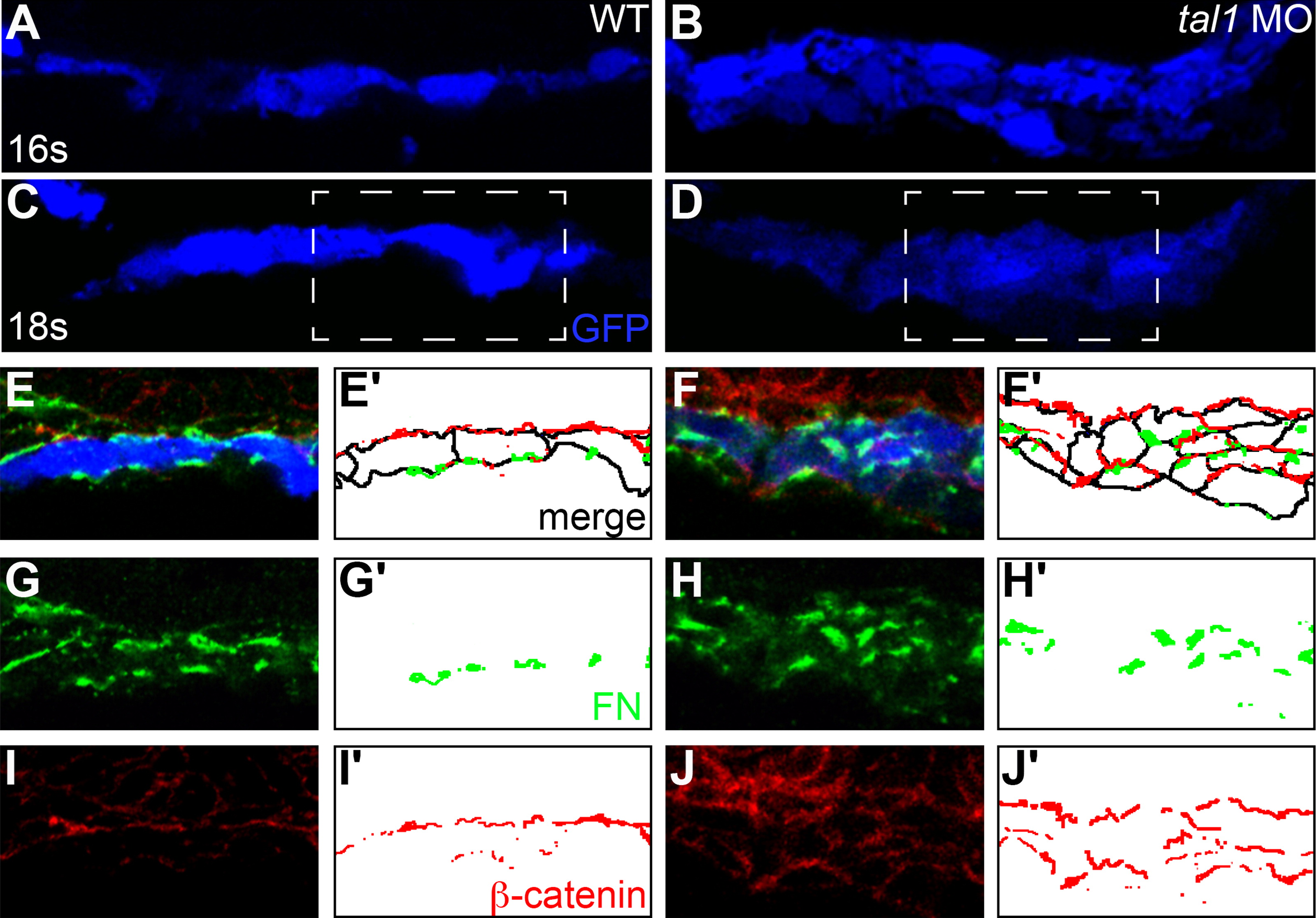
Fig. 4 The architecture of the early endocardium is disrupted in tal1-deficient embryos. (A?J) Transverse sections of wild-type (A, C, E, G, and I) and tal1-deficient embryos (B, D, F, H, and J), dorsal to the top, at 16s (A and B) and 18s (C?J). Immunofluorescence detects localization of fibronectin (FN; green) (E?H) and β-catenin (red) (E, F, I, and J); in addition, an anti-GFP antibody allows visualization of Tg(kdrl:GRCFP) expression (blue) in the endocardium (A?F). Together, these markers highlight the shapes and arrangement of the endocardial cells. Endocardial cells form a single-layered sheet of cells at 16s (A), after they have migrated to the midline, and this sheet of cells is maintained at 18s (C). In tal1-deficient embryos, endocardial cells stack on top of each other, forming a multi-layered cluster (B and D). The boxed areas in (C and D) are highlighted in (E?J). The apical-basal polarity of wild-type endocardial cells (E) is indicated by basal deposition of FN (G) and apical enrichment of β-catenin (I). In tal1-deficient embryos (F), localization of FN (H) and β-catenin (J) highlight the dysmorphic endocardial architecture and aberrant cell shapes. (E′?J′) Cartoons indicate regions of FN (green) and β-catenin (red) localization within the endocardium. Endocardial cell outlines are marked in black. The FN and β-catenin that are presumed to be located within the endoderm that overlies the endocardium are excluded from the cartoons.
Reprinted from Developmental Biology, 383(2), Schumacher, J.A., Bloomekatz, J., Garavito-Aguilar, Z.V., and Yelon, D., tal1 regulates the formation of intercellular junctions and the maintenance of identity in the endocardium, 214-226, Copyright (2013) with permission from Elsevier. Full text @ Dev. Biol.