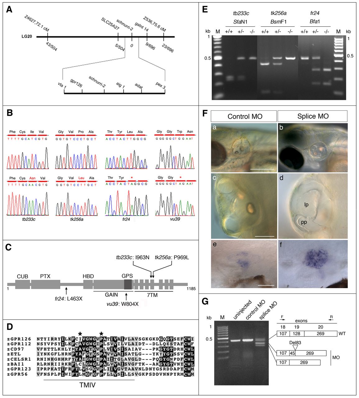
Fig. 5 Positional cloning, identification and confirmation of mutations in gpr126. (A) Genetic map of the lau locus. Numbers of meiotic recombinants for the flanking SSLP markers and SNP markers in slc25a27, schnurri2 and galnt14 are shown. (B) Sequence analysis of gpr126 cDNA in wild type (upper panels) and lau mutants (lower panels), with predicted changes to the coding sequence. (C) Schematic diagram of the Gpr126 protein, with its conserved domains: CUB (Complement C1r/C1s, Uegf, BMP1), PTX (Pentraxin), HBD (hormone binding domain), GAIN (GPCR autoproteolysis inducing) domain, GPS (GPCR proteolytic site) motif and 7TM (7-transmembrane) domain (not to scale). Positions of the mutations are shown. (D) Amino acid comparison of the fourth transmembrane (TMIV) region from eight zebrafish adhesion class GPCRs, showing the conserved hydrophobic residue at position 963 (mutated in tb233c), and the highly conserved proline (P) at position 969 (mutated in tk256a) (asterisks). (E) Genotyping of lau mutant fish by restriction digest of PCR-amplified genomic DNA. In tb233c, the mutation eliminated an SfaN1 site; in tk256a, the mutation eliminated a BsmF1 site; in the fr24 allele, a Bfa1 site was gained. (F,G) gpr126 morpholino injection recapitulates the lau mutant ear phenotype. (F) Wild-type (nac) embryos (left hand panels) injected with 5 ng control morpholino exhibit normal ear morphology at 5 dpf (a,c), and low vcanb expression at 5 dpf (e). Wild-type (nac) embryos injected with 5 ng gpr126 morpholino (right hand panels) have abnormal projection outgrowth (b,d). The lateral projection (lp) is enlarged, and the posterior projection (pp) in this ear has grown past the lateral projection without fusing. Expression of vcanb is upregulated (f). (G) RT-PCR analysis of gpr126 mRNA processing of the 19th exon in gpr126 morpholino-injected, or in control (mismatched) morpholino-injected, embryos at 5 dpf. Sequencing confirmed two aberrant splice variants. Panels c,d are dorsal views, anterior towards the top. Scale bars: 200 μm in a,b; 50 μm in c,d; 50 μm in e,f.
Image
Figure Caption
Figure Data
Acknowledgments
This image is the copyrighted work of the attributed author or publisher, and
ZFIN has permission only to display this image to its users.
Additional permissions should be obtained from the applicable author or publisher of the image.
Full text @ Development

