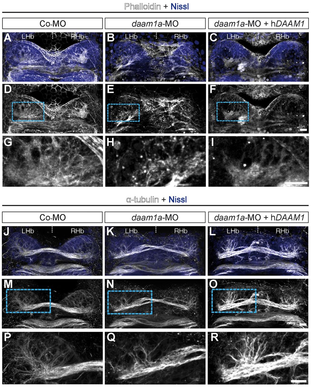
Fig. 5 Knockdown of Daam1a affects the organisation of filamentous actin and microtubules in the Hb. (A,D,G) The Hb of control embryos labelled with fluorescent phalloidin (F-actin, white) and counterstained with Nissl (to delineate the cellular context, purple) show a distinct smooth fibrillar organisation of F-actin, with increased compaction in the neuropil domains. (B,E,H) The Hb of daam1a-MO embryos show an irregular pattern of F-actin, with shorter and thicker bundles. (C,F,I) The Hb of embryos co-injected with daam1a-MO and hDAAM1, show a smooth fibrillar pattern of F-actin similar to controls. (J,M,P) The Hb of control embryos immunostained against α-tubulin (white) and counterstained with Nissl showed a distinct asymmetric distribution of α-tubulin corresponding to the neuropil domains. (K,N,Q) The Hb of daam1a-MO embryos show decreased and less elaborate α-tubulin staining. (L,O,R) The Hb of embryos co-injected with daam1a-MO and hDAAM1, show increased levels and thickness of α-tubulin (+) bundles than controls. G-I and P-R correspond to magnification views of the boxed regions in D-F and M-O, respectively. Images correspond to dorsal views of maximum intensity z-stack confocal projections, with anterior to the top. LHb, left Hb; RHb, right Hb. Scale bars: 20 μm.
Image
Figure Caption
Figure Data
Acknowledgments
This image is the copyrighted work of the attributed author or publisher, and
ZFIN has permission only to display this image to its users.
Additional permissions should be obtained from the applicable author or publisher of the image.
Full text @ Development

