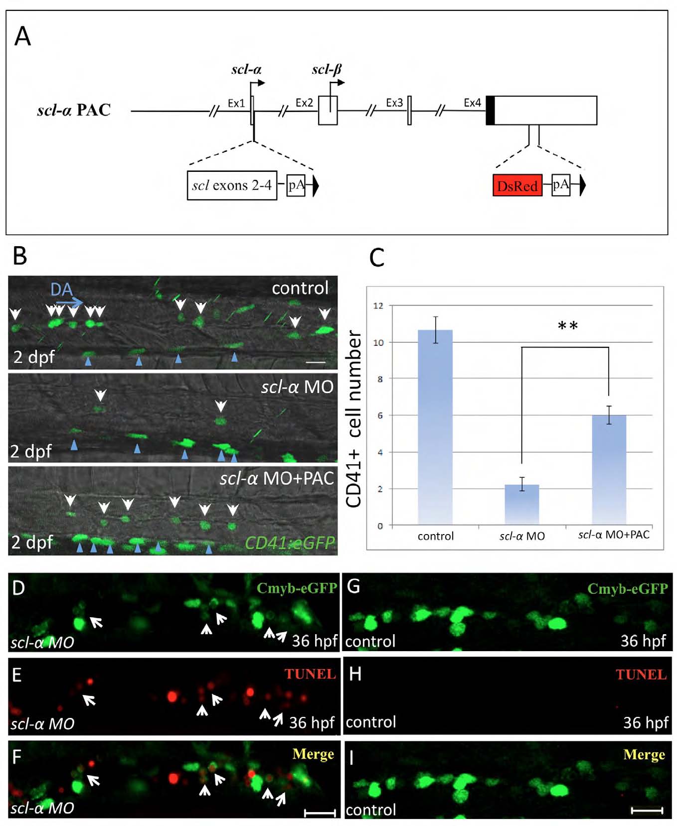
Fig. S5 scl-α MO is specific to the defects of HSC maintenance in the AGM of scl-α morphants. (A-C) The loss of HSCs is partially rescued in the scl-α morphant receiving scl-α PAC expression. (A) The scl-α PAC used for rescue. DsRed and an SV40 polyadenylation signal were inserted in exon 4 to interrupt the transcription of scl-β. To introduce normal expression of scl-α, the DNA sequences of scl exon 2, 3, 4 and an SV40 polyadenylation signal were inserted behind exon 1. (B) CD41:eGFP+ HSCs in the AGM region of live 2 dpf control embryos, scl-α morphants and scl-α morphants injected with scl-α PAC. White arrows indicate CD41:eGFP+ HSCs in the AGM. Blue arrowheads identify pronephric duct cells. Scale bar: 20 μm. (C) Statistical analysis showing the number of CD41:eGFP+ HSCs (per five somites) in the AGM of 2 dpf control embryos, scl-α morphants and scl-α morphants injected with scl-α PAC. (D-I) Double immunostaining of Cmyb-eGFP and TUNEL in the AGM of 36 hpf scl-α morphants (D-F) and control embryos (G-I). White arrows indicate three Cmyb:eGFP+ cells undergoing apoptosis in the AGM. Scale bar: 20 μm.
Image
Figure Caption
Acknowledgments
This image is the copyrighted work of the attributed author or publisher, and
ZFIN has permission only to display this image to its users.
Additional permissions should be obtained from the applicable author or publisher of the image.
Full text @ Development

