Fig. 3
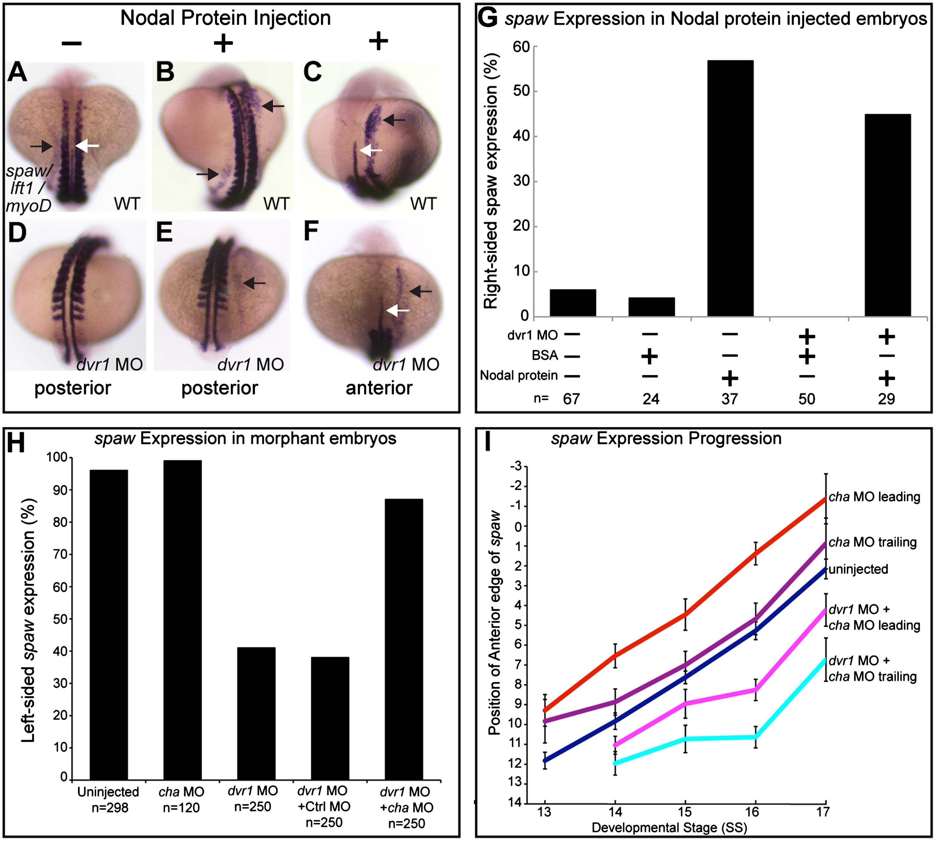
Initiation in LPM can occur in dvr1 morphants and timing is dependent on mix of activators and inhibitors. (A) Control (BSA injected) wildtype embryos show normal left-sided spaw expression. Embryos injected with Nodal protein on right side, (B) anterior view and (C) same embryo, posterior view, had both native left-sided and ectopic right-sided LPM spaw expression (black arrows). Midline lft1 expression (white arrow) and somitic myoD expression serve as spatial landmarks. (D) Control (BSA injected) dvr1 morphant embryos exhibit no spaw expression. (E) and (F) dvr1 morphant embryos injected with Nodal protein on the right side exhibit ectopic right-sided spaw expression. (E) Same Nodal-injected dvr1 morphant, (E) posterior and (F) anterior views. (G) Quantification of right-sided spaw expression in uninjected controls, BSA injected controls, Nodal protein injected into right side of wildtype embryos, dvr1 morphants injected with BSA on the right side, and dvr1 morphants injected with Nodal protein on the right side. (H) Quantification of left-sided (normal) spaw expression in single and double morpholino injected embryos. (I) Quantitative measurements of initiation and posterior-to-anterior propagation of spaw expression in LPM of wildtype, cha morphants, and dvr1+cha double morphants.
Reprinted from Developmental Biology, 382(1), Peterson, A.G., Wang, X., and Yost, H. J., Dvr1 transfers left-right asymmetric signals from Kupffer's vesicle to lateral plate mesoderm in zebrafish, 198-208, Copyright (2013) with permission from Elsevier. Full text @ Dev. Biol.

