
|
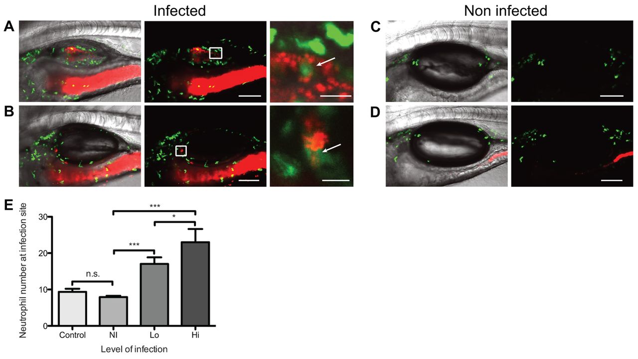
Fig. 5
Neutrophils are present at increased levels in swimbladder infection. (A?D) Cohorts of 20 mpx:GFP fish were infected by immersion with C. albicans CAF2-dTomato and imaged by confocal microscopy at 5 dpi. (A,B) Neutrophil accumulation at the site of infection. (A) High-level infection in non-inflated swimbladder; (B) low-level infection in fully inflated swimbladder. Panel on the right is a magnification of red and green channels (white box) showing direct contact between neutrophil and C. albicans (arrows). Animated z-stack of panel A, right, is shown in supplementary material Movie 8. Animated z-stack of panel B, right, is shown in supplementary material Movie 7. (C,D) Non-infected control fish. (C) C. albicans was not added to the media and (D) C. albicans immersion, no infection. (E) Number of neutrophils in the swimbladder per individual fish. Average and standard error of three independent experiments are shown (pooled data). One-way ANOVA and Bonferroni post-hoc test; ***P<0.001; *P<0.05; n.s. non significant. Scale bars: 100 μm (left panel) and 20 μm (right panel) for magnification. Maximum projections of n slices: n=7 (A?D) and n=5 for magnification of A and B. Images are representative of four independent experiments.