Fig. 5
Fig. 5
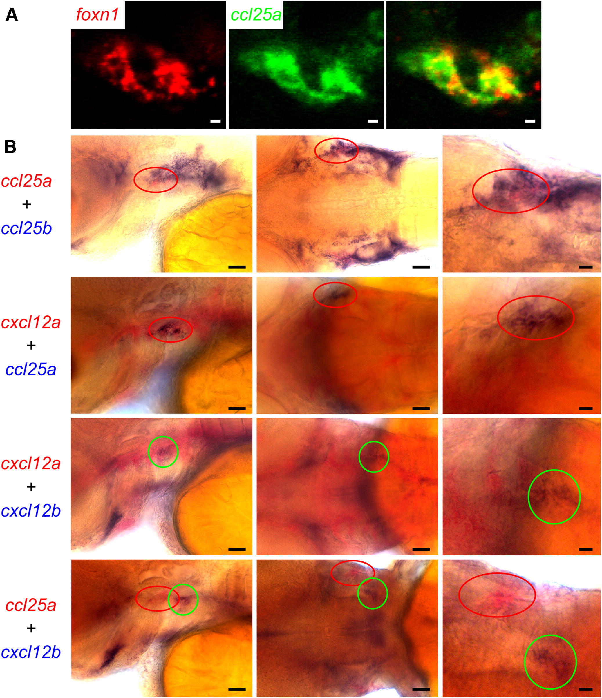
Expression Domains of ccl25 and cxcl12 Chemokines in 3 dpf Embryos
(A) Whole-mount double-fluorescence RNA in situ hybridization of foxn1 (red fluorescence) and ccl25a (green fluorescence). The colocalization in thymic epithelia of foxn1 and ccl25a was revealed by confocal fluorescence microscopy. Representative results for <50 embryos. Scale bars represent 5 μm.
(B) High-resolution double-colorimetric RNA in situ hybridization for the indicated genes; color code corresponds to the images shown in lateral (first column), dorsal (second column), and high-power (third column) views. Expression of ccl25a marks the thymic epithelium (see A). The red and green circles mark the thymus and the parathymic regions, respectively. Representative results for <50 embryos. Scale bars represent 50 μm for overviews, 20 μm for high-power views. See also Figure S3.
Reprinted from Immunity, 36(2), Hess, I., and Boehm, T., Intravital Imaging of Thymopoiesis Reveals Dynamic Lympho-Epithelial Interactions, 298-309, Copyright (2012) with permission from Elsevier. Full text @ Immunity

