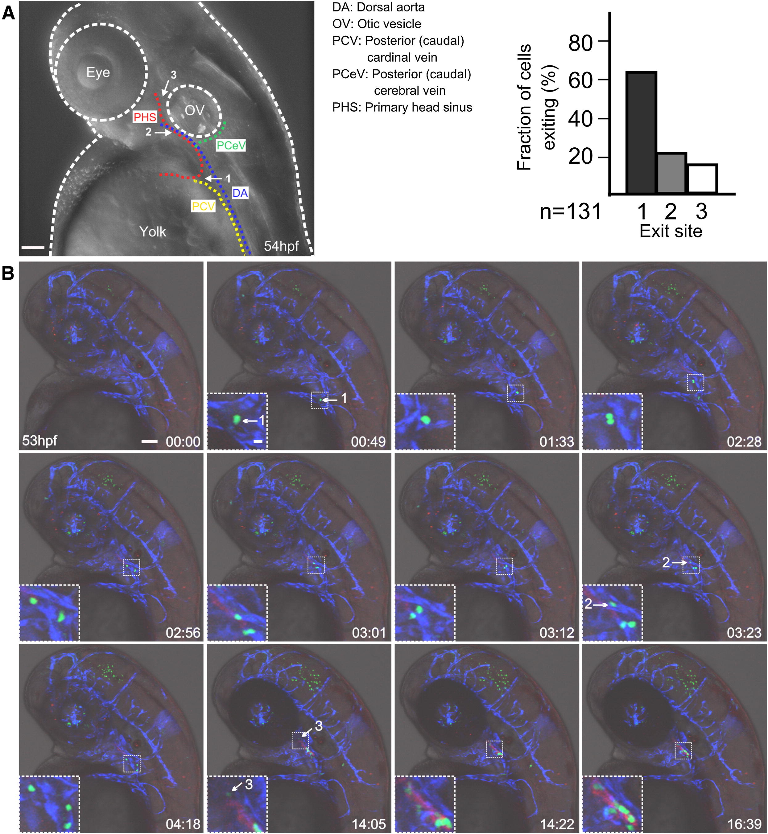
Fig. 3
Three Major Routes of Thymus Colonization
(A) Schematic highlighting major anatomical structures relevant to the three major sites of extravasation (numbered in order of importance) of lymphocyte progenitors. The distribution of 131 exit events (recorded from 15 embryos for the 52?68 hpf observation period) among the three sites is shown on the right.
(B) Still photographs of a time-lapse recording of a representative triple-transgenic embryo (ikaros:eGFP [green fluorescence]; foxn1:mCherry [red fluorescence]; flk1:CFP [blue fluorescence]). The still series begins at 53 hpf (00 hr:00 min). The sites of extravasation from vessels are numbered as in (A); lymphocyte progenitors exhibit bright green fluorescence. The marked regions are displayed in higher magnifications in the insets. For details, see text. Note that some neurons in the retina and the brain also express ikaros; a subset of skin cells also expresses foxn1. Representative results for ten embryos. Scale bars represent 50 μm for overviews, 5 μm for insets.
See also Figure S2 and Movie S4.
Reprinted from Immunity, 36(2), Hess, I., and Boehm, T., Intravital Imaging of Thymopoiesis Reveals Dynamic Lympho-Epithelial Interactions, 298-309, Copyright (2012) with permission from Elsevier. Full text @ Immunity

