Fig. S2
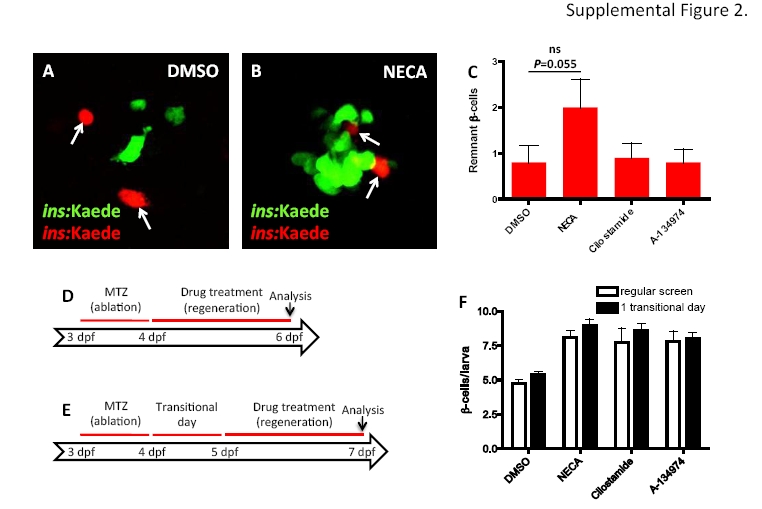
Survival of β cells does not significantly contribute to increased regeneration in the presence of the hit-compounds, related to Figure 2.
(A-C) As described for Figure 2, we examined β cell survival by photo-converting the fluorescent protein Kaede. At 3 dpf, before ablating the β cells with MTZ between 3-4 dpf, we converted Tg(ins:Kaede)-expressing β cells from green to red by exposing them to UV-light. After two days of regeneration (6 dpf), the surviving β cells are red and green (yellow overlap), whereas the newly formed β cells are green-only. (A-B) We observed β cells that had not yet been ablated and cleared by 6 dpf; however, these cells do not have an active insulin promoter and are therefore red-only (arrows). Because these cells do not seem to be functional they were not included in the quantification presented in Figure 2. (C) Quantification of red-only remnant β cells showed no significant difference between the different treatments, although there was a trend for an increased number of remnant β cells after NECA treatment. n = 10 larvae for each group.(D-F) β cell regeneration was examined with or without a transitional day that allowed β cell ablation to conclude and MTZ to wash out before drug treatment commenced. (D) Regular schema for the β cell regeneration screen. (E) Schema for examining β cell regeneration following a transitional day. (F) Quantification of β cell regeneration using wide-field fluorescence microscopy showing that NECA, Cilostamide, and A-134974 all increased regeneration with equal potency in the regular schema as in the schema with a transitional day. n = 38-66 larvae for each group. Error bars represent SEM.
Reprinted from Cell Metabolism, 15(6), Andersson, O., Adams, B.A., Yoo, D., Ellis, G.C., Gut, P., Anderson, R.M., German, M.S., and Stainier, D.Y., Adenosine Signaling Promotes Regeneration of Pancreatic beta Cells In Vivo, 885-894, Copyright (2012) with permission from Elsevier. Full text @ Cell Metab.

