
|
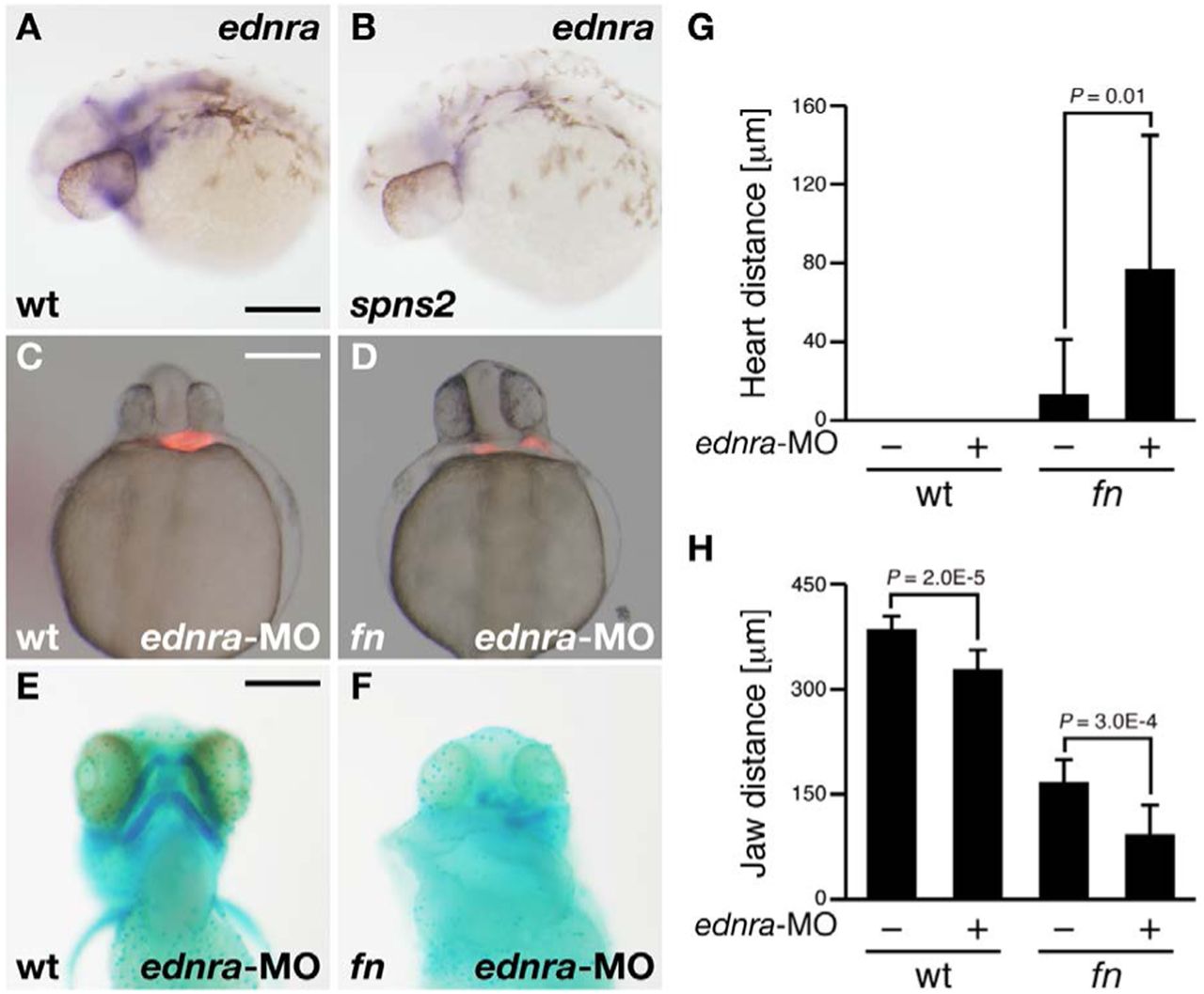
Fig. 4
Knockdown phenotype of endothelin receptor A (ednra) in fn mutants.
(A,B) Whole-mount in situ hybridization using the ednra RNA probe. The expression of ednra was suppressed in the spns2 mutant. Both images show lateral views at 30hpf. (C,D) Cardiac morphology visualized by mRFP expression derived from Tg(cmlc2:mRFP). Both images show ventral views at 28hpf. (E,F) Lower jaw morphology at 4dpf was visualized by Alcian Blue staining (ventral view). Genotyping was performed by genomic sequencing after taking pictures. wt (A,C,E), spns2 mutant (B) and fn mutant (D,F). Scale bars: 200μm. (G,H) Average distances between hearts (G) and anterior?posterior distances of the ventral pharyngeal arch (H) from multiple experiments; error bars represent standard deviations.