Fig. 7
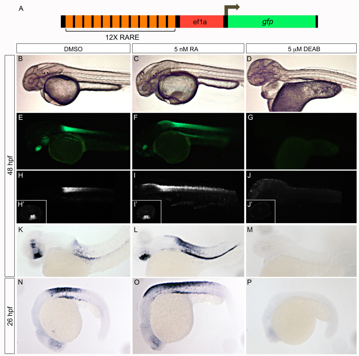
Validation of RARE:eGFP reporter. Tg(12xRARE-ef1a:eGFP)sk71 transgenic embryos (described in A) were evaluated for altered signals when RA signaling was manipulated. Embryos were treated with a vehicle control, DMSO (B, E, H, H′, K, N), 5 nM all-trans retinoic acid (C, F, I, I′, L, O), or 5 μM DEAB (D, G, J, J′, M, P). We note an expansion of hindbrain/spinal cord expression of the transgene in 5 nM RA-treated embryos, and a complete loss in 5 μM DEAB-treated embryos. Altered expression is detectable by examination of fluorescence using a stereomicroscope (B-G), confocal microscope (H-J′), or by in situ hybridization with a probe to eGFP(K-P). All embryos are shown in lateral view at 48 hpf (B-M) or 26 hpf (N-P). The inset panels (H′-J′) display reporter activity in the ventral retina.

