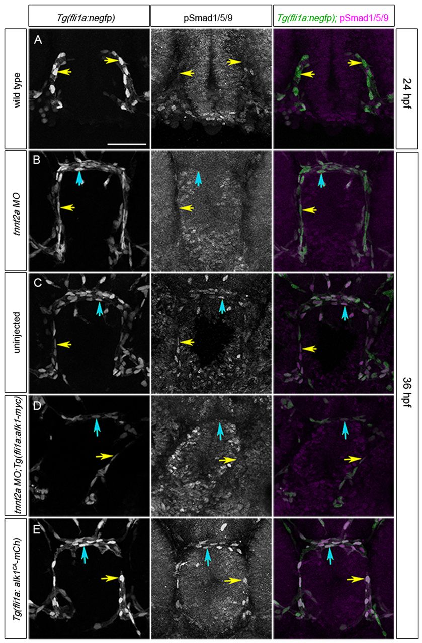
Fig. 2
Alk1 activity is dependent on blood flow. (A-E) pSmad1/5/9 (middle column) in endothelial cell nuclei (marked by fli1a:negfp transgene, left column); in merge (right column), EGFP-expressing endothelial cell nuclei are green and pSmad1/5/9 immunofluorescence is magenta. (A) 24 hpf wild type, prior to blood flow; (B) 36 hpf tnnt2a morphant (no flow); (C) 36 hpf wild type; (D) 36 hpf tnnt2a morphant harboring a fli1a:alk1-myc transgene; (E) 36 hpf tnnt2a morphant harboring a fli1a:alk1CA-mCh transgene. Tg(fli1a:alk1CA-mCh) embryos do not have lumenized vessels. In merge (right column), EGFP-expressing endothelial cell nuclei are green, pSmad1/5/9 immunofluorescence is magenta. Yellow and blue arrows indicate endothelial cells in the caudal division of the internal carotid artery (CaDI) and basal communicating artery (BCA), respectively. 2D confocal projections of 50 μm frontal sections, dorsal upwards. Scale bar: 50 μm. See supplementary material Table S1 for fluorescence quantitation.

