
|
Fig. 5
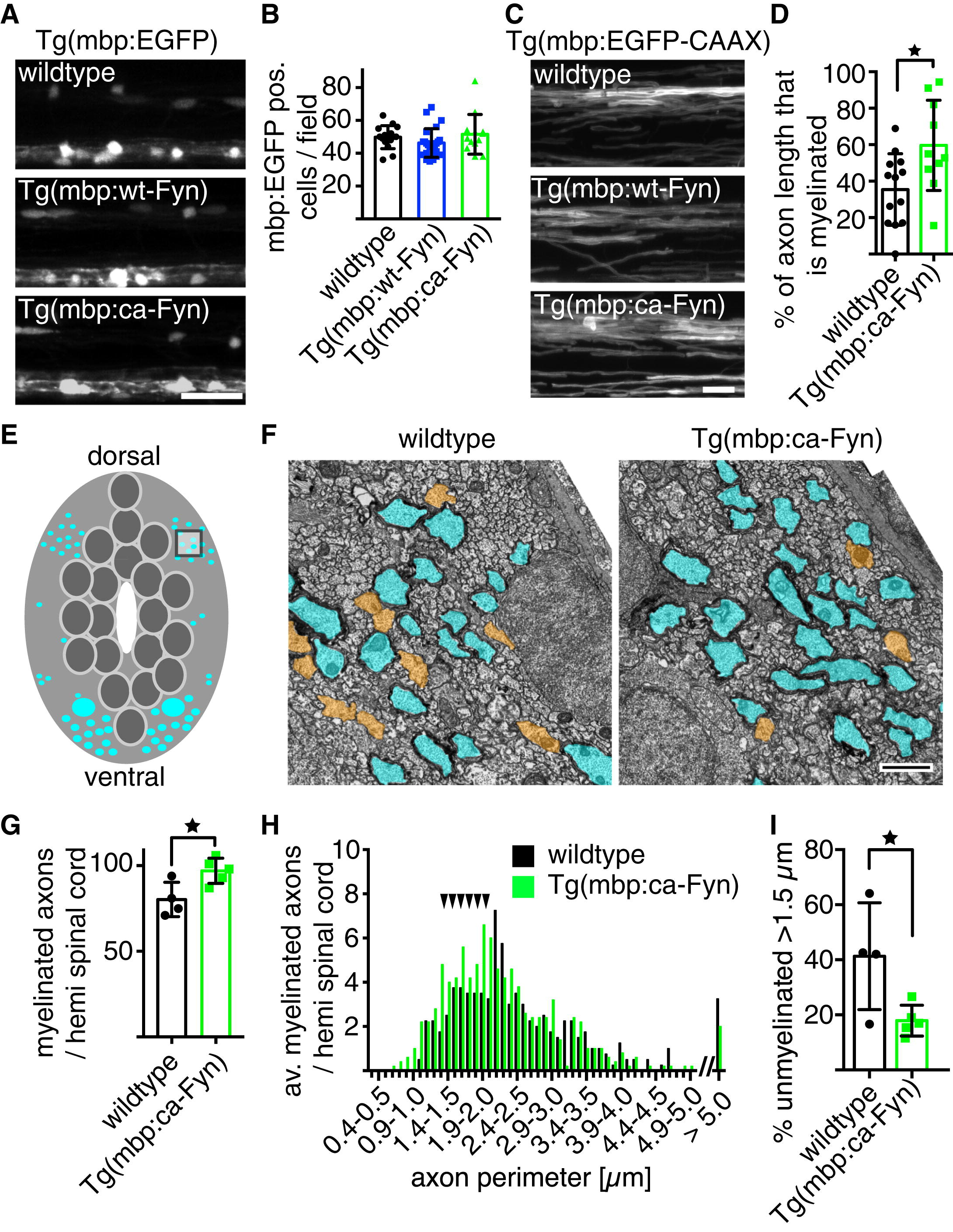
Constitutive Activation of Fyn in Myelinating Oligodendrocytes Causes Precocious Myelination without Affecting Cell Number (A) Confocal images of Tg(mbp:EGFP) wild-type control (top), Tg(mbp:wt-Fyn) (middle), and Tg(mbp:ca-Fyn) (bottom) animals at 8 dpf show no difference in oligodendrocyte number or distribution. Scale bar, 25 μm. (B) Quantification of myelinating oligodendrocyte number at 8 dpf reveals no differences between wild-type, Tg(mbp:wt-Fyn), and Tg(mbp:ca-Fyn) animals. Significance was assessed using one-way ANOVA. Error bars indicate SD. (C) High-magnification views of the dorsal spinal cord at 8 dpf in Tg(mbp:EGFP-CAAX) control (top), Tg(mbp:wt-Fyn) (middle), and Tg(mbp:ca-Fyn) (bottom) animals show that there are additional myelinated axons in Tg(mbp:ca-Fyn) animals. Scale bar, 10 μm. (D) Quantification showing percentage of single axon length myelinated in control compared to Tg(mbp:ca-Fyn) animals by 5 dpf. Significance was assessed using Student?s two-tailed unpaired t test. Error bars indicate SD. (E) Schematic transverse cross-section of the larval zebrafish spinal cord indicating myelinated axons in cyan. (F) Transmission electron microscope images of transverse sections of spinal cords at 8 dpf of an area indicated in (E) in wild-type (left) and Tg(mbp:ca-Fyn) (right) animals. There is a larger number of myelinated axons (shaded in cyan) in Tg(mbp:ca-Fyn) animals and a smaller number of large caliber (>1.5μm perimeter) unmyelinated axons (shaded in orange). Scale bar, 1 μm. (G) The number of myelinated axons per hemi spinal cord is increased in Tg(mbp:ca-Fyn) animals compared to wild-type. Significance was assessed using Student?s two-tailed t test. Error bars indicate SD. (H) The distribution of myelinated axon size (assessed by axonal perimeter) in wild-type and Tg(mbp:ca-Fyn) animals is similar. The additional axons myelinated in Tg(mbp:ca-Fyn) animals compared to control are almost all between 1 and 2 μm in perimeter (arrowheads). (I) Quantification of percentage unmyelinated axons with a perimeter >1.5 μm in wild-type and Tg(mbp:ca-Fyn) dorsal spinal cord. Significance was assessed using Student?s two-tailed unpaired t test. Error bars indicate SD. See also Figure S1.
Reprinted from Developmental Cell, 25(6), Czopka, T., Ffrench-Constant, C., and Lyons, D.A., Individual Oligodendrocytes Have Only a Few Hours in which to Generate New Myelin Sheaths In Vivo, 599-609, Copyright (2013) with permission from Elsevier. Full text @ Dev. Cell