
|
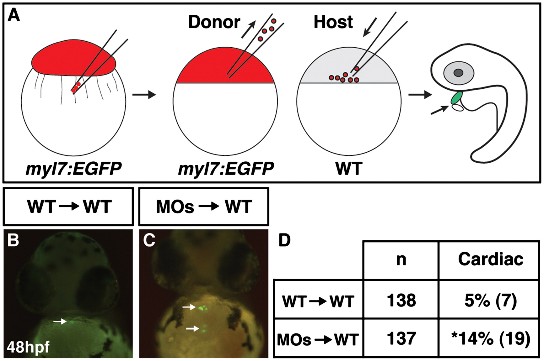
Fig. 6
Tcf7l1s cell autonomously promote CM development. (A) Schematic of the cell transplantation strategy used for assessing cell autonomy. Red indicates injection with the rhodamine-dextran lineage tracer. (B) Representative image of a CM (arrow) that contributed to the heart from a WT/Tg(5.1myl7:EGFP) donor cell transplant into a WT host embryo. (C) Representative image of CMs (arrows) that contributed to the heart from a Tcf7l1 deficient/Tg(5.1myl7:EGFP) donor cell transplanted into a WT host. We found an increase in the frequency of CMs that contribute to the heart from Tcf7l1 deficient/Tg(5.1myl7:EGFP) donor cells and that these cells formed large clusters of CMs (Supplemental Table 1). (B) and (C) are frontal views. (D) Frequency of contribution to the heart from donor cells in transplantation experiments. Asterisk indicates a statistically significant difference compared to control transplant experiments.
Reprinted from Developmental Biology, 380(2), Sorrell, M.R., Dohn, T.E., D'Aniello, E., and Waxman, J.S., Tcf7l1 proteins cell autonomously restrict cardiomyocyte and promote endothelial specification in zebrafish, 199-210, Copyright (2013) with permission from Elsevier. Full text @ Dev. Biol.