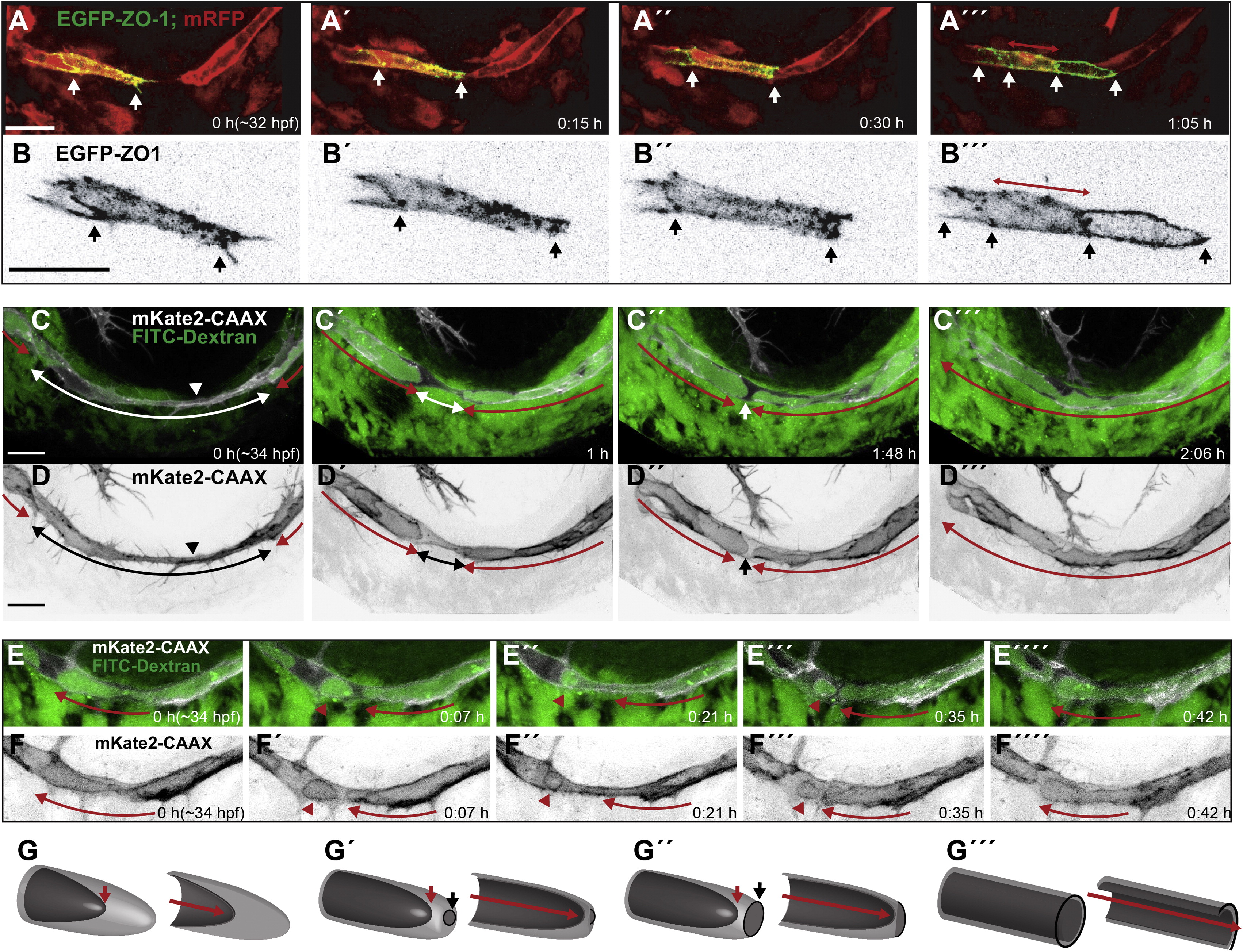
Fig. 2
Lumen Formation in the PLA (A and B) Still pictures of a time-lapse movie showing contact and lumen formation in a transgenic embryo Tg(fliep:GFF)ubs3,(UAS:mRFP),(UAS:EGFP-ZO-1)ubs5. One of the tip cells is labeled with EGFP-ZO-1 (green). ECs cytoplasm is red. White arrows point the ends of the labeled cell (A–A3) and the edges of EGFP-ZO-1 rings (A32) when the cell becomes a unicellular tube with transcellular lumen (A32, red arrow). (B) shows the EGFP-ZO-1 labeled cell alone, enlarged 2× (black). (C and D) Still pictures of a time-lapse movie showing lumen formation in the PLA in a transgenic embryo TgBAC(kdrl:mKate-CAAX)ubs16 injected with FITC-dextran. After the tip cell contact is established, the luminal/apical membrane (red arrows) invaginates toward the contact site (C, D, arrowheads). White/black arrows shows the nonlumenized cell parts. Dextran (green) reaches the end of the luminal membrane, showing that the lumen is connected to the circulation. The membrane invagination advances from both sides (red arrows) until the two lumens are only separated by a small part of the cell body (C3/D3 white/black arrows). Perfusion of the lumen involves membrane fusion and connects the two lumens allowing the dextran to flow through. (E and F) Still pictures of a time-lapse movie showing possible apical membrane rearrangements after initial lumen perfusion. A fully formed lumen (red arrow marks continuous lumen) partially collapses leading to separation of an apical membrane compartment in the form of a large sphere filled with Dextran (red arrowheads). The sphere can be maintained for a certain time (E3 and E32), and finally it reconnects with the neighboring luminal compartment. (G) A 3D model of a tip cell undergoing transcellular lumen formation through cell membrane invagination. The basal membrane is light gray; the apical/luminal membrane is dark gray. Red arrows point out the lumen end and the lumen length (in the cross section models). Black arrows show the new contact formation site, with junctions marked in black and new apical membrane in dark gray within. Scale bars, 20 μm. See also Figures S1 and S2 and Movies S2 and S3.
Reprinted from Developmental Cell, 25(5), Lenard, A., Ellertsdottir, E., Herwig, L., Krudewig, A., Sauteur, L., Belting, H.G., and Affolter, M., In Vivo analysis reveals a highly stereotypic morphogenetic pathway of vascular anastomosis, 492-506, Copyright (2013) with permission from Elsevier. Full text @ Dev. Cell

