
|
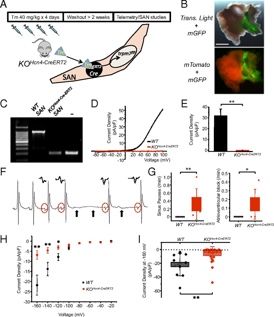
Fig. 8
Postnatal SAN/AVN restricted Trpm7 deletion recapitulates the phenotype of global cardiac Trpm7 KO. (A) SAN-restricted Trpm7 deletion (KOHcn4-CreERT2) was achieved in Hcn4-CreERT2 � Trpm7fl/fl/Trpm7fl/- mice at 6?8 wk of age by treating with tamoxifen (40 mg/kg) by gavage for 4 d, followed by >2-wk washout period before telemetry and SAN studies (B) (Upper) Transmitted light + GFP fluorescence image of Hcn4-CreERT2-ROSA26mTmG right atrium after tamoxifen treatment. (Lower) Fluorescence mTomato/mGFP image showing recombination (mGFP) restricted to SAN region. (Scale bar, 1 mm.) (C) PCR across exon 17 from genomic DNA isolated from dissected KOHcn4-CreERT2 and WT SAN. Tail DNA from Trpm7fl/- serves as a positive control for Trpm7 exon 17 deletion (-). (D) Representative TRPM7 current?voltage traces measured in WT SAN and KOHcn4-CreERT2 SAN. (E) Mean TRPM7 current density in WT SAN and KOHcn4-CreERT2 SAN. (F) Representative ECG showing an episode of SP observed in a telemetered KOHcn4-CreERT2 mouse. Solid arrows denote location of expected p waves. Red dashed circles and Insets above show change in p-wave morphology after SP, indicative of ectopic atrial focus. (G) Box plots with overlying data points showing the distribution of the frequency SPs (Left) and AVB (Right) observed over 24 h of telemetric monitoring in WT (n = 7) and KOHcn4-CreERT2 (n = 5) mice. (H) If current?voltage relationship from WT SAN (n = 11) compared with KOHcn4-CreERT2 SAN (n = 25). (I) Box plots with overlying data points showing the mean and distribution of If current densities at 160 mV from WT SAN (n = 11) compared with KOHcn4-CreERT2 SAN (n = 25). *P < 0.05, **P < 0.01.