
|
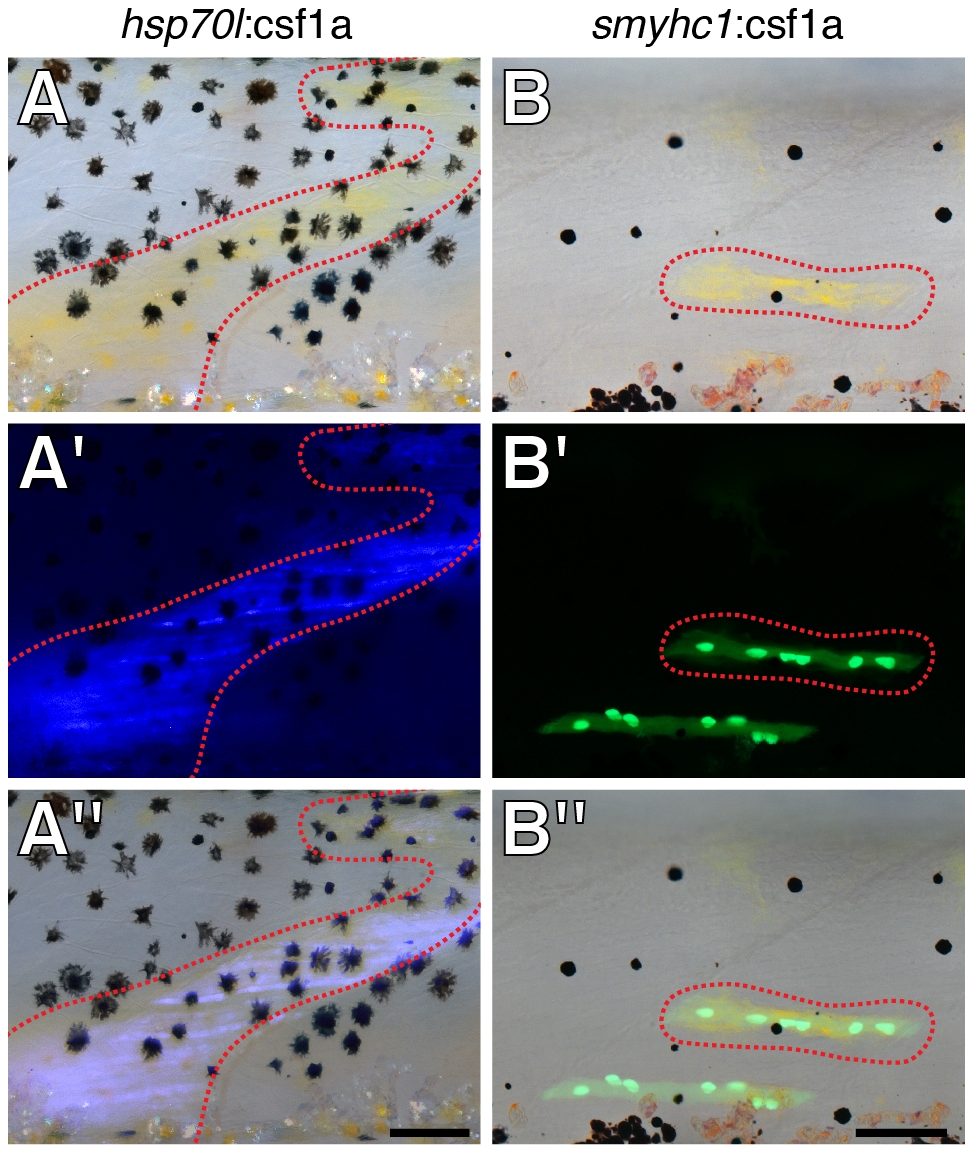
Fig. 7
Localized Csf1 expression directed xanthophore development.
(A) Ectopic xanthophores (red dashed line) developed over the dorsal myotome in association with Csf1a-expressing cells transplanted from a wild-type, Tg(hsp70l:csf1a-IRES-nlsCFP) donor to a bnc2 mutant host. Larva shown at 7.9 SSL. (A2) Nuclear CFP expression in the myotome. (A3) Merge. (B) Ectopic xanthophores in a wild-type larva developed over the dorsal myotome in association with a slow muscle fiber of the myotome expressing Csf1a from plasmid smyhc1:TetGBD-TREtightBactinTRX:nlsVenus-V2a-c sf1a.Larva shown at 7.5 SSL. (B2) Nuclear Venus expression. (B3) Merge. (Sample sizes: hsp70l, n = 8; smyhc1, n = 10.) Scale bars: in (A3) 100 μm for (A); in (B) 100 μm for (B).