Fig. 5
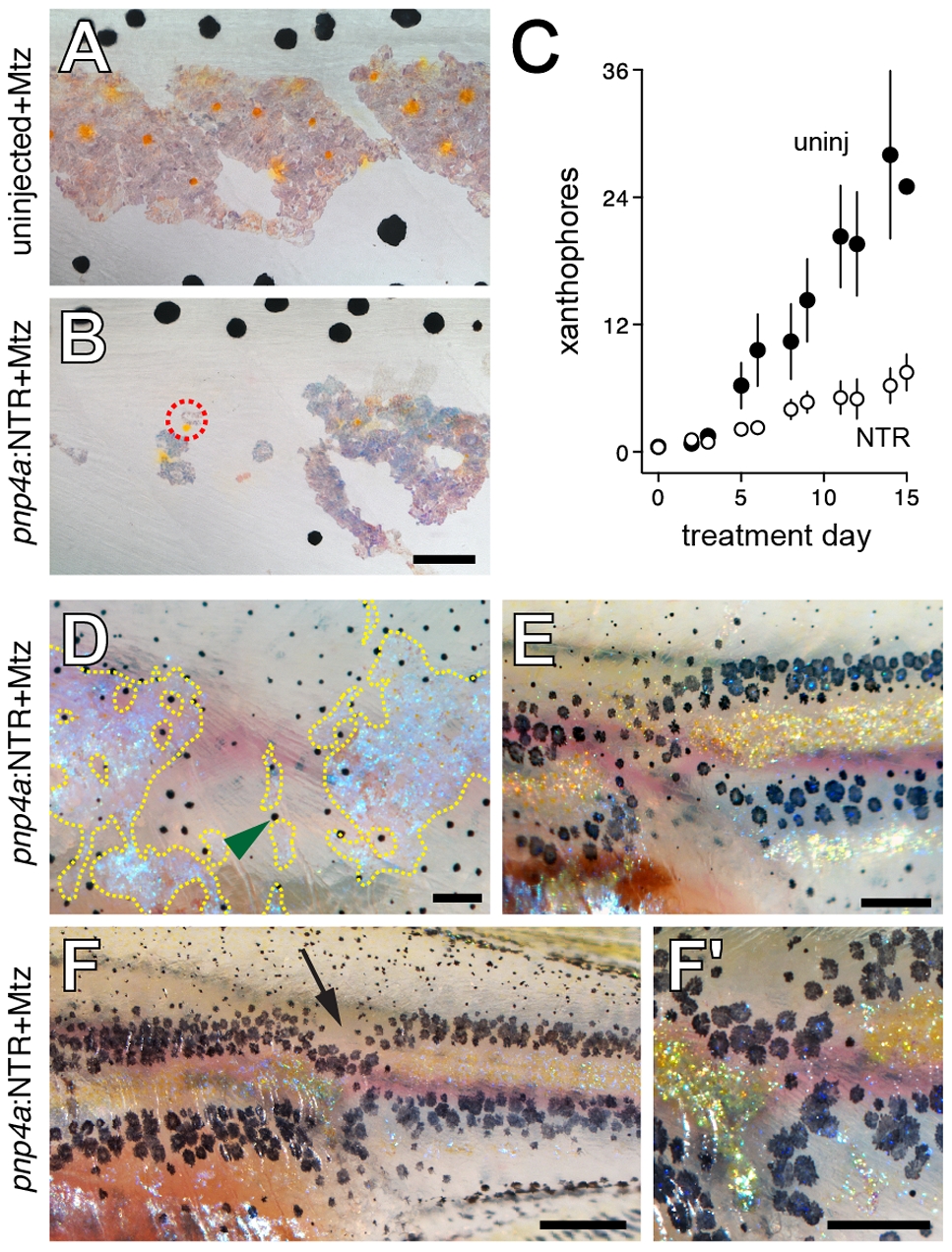
Iridophore ablation perturbed xanthophore and melanophore patterning.
(A, B) Wild-type siblings that were either not injected (A) or injected (B) with pnp4a:NTR plasmid and then treated with Mtz beginning at 5 SSL, prior to the the onset of xanthophore differentiation. Controls (A) exhibited normal interstripe iridophores and xanthophores whereas iridophore-ablated individuals developed xanthophores primarily in association with residual iridophores (e.g., dashed red circle in B). (C) Numbers of xanthophores (meansąSE) in stage-matched siblings treated with Mtz that were either uninjected or injected with pnp4a:NTR plasmid. Xanthophore numbers did not differ between groups at the onset of the experiment but iridophore-ablated individuals showed an increasingly severe xanthophore deficiency compared to uninjected larvae as the experiment proceeded (genotype×day interaction, F1,10 = 2.7, P<0.005; initial sample sizes: uninjected, n = 13; pnp4:NTR, n = 13). During later development, new xanthophores ultimately developed more broadly over the flank and in association with regenerating iridophores; iridophore ablations after xanthophores had differentiated typically did not affect these cells (not shown). (D?F) Examples of larvae (9.5 SSL) exhibiting melanophore patterning defects following earlier iridophore ablations (started at 6.0 SSL). Melanophores have colonized regions from which iridophores were ablated, though a few regenerative or persisting iridophore remained. In the lighting used here, iridophores are blue or gold iridescent. (D) Melanophores occupy a region from which iridophores were ablated (residual or regenerated iridophores outlined by dashed yellow lines). Green arrowhead, one of several melanophores localized adjacent to remaining iridophores. Fish shown in A, C and D were treated with epinephrine prior to imaging. (E) Melanophore stripes are broken at site of iridophore ablation and melanophores appear to ?wrap around? residual interstripe iridophores on either side of the ablation. (F) In another individual, melanophores stripes are constricted where iridophores have been ablated (arrow). Close-up in F2. Fish in E and F were not treated with epinephrine, so that melanin reveals peripheral processes of melanophores. Most small melanophores in dorsal regions are associated with developing scales and will not contribute to the stripe pattern [45]. (Total sample size, N = 40.) Scale bars: in (B) 60 μm for (A,B); in (D) 200 μm for (D); in (E) 500 μm for (E); in (F) 100 μm for (F); in (F2) 60 μm for (F2).

