Fig. 8
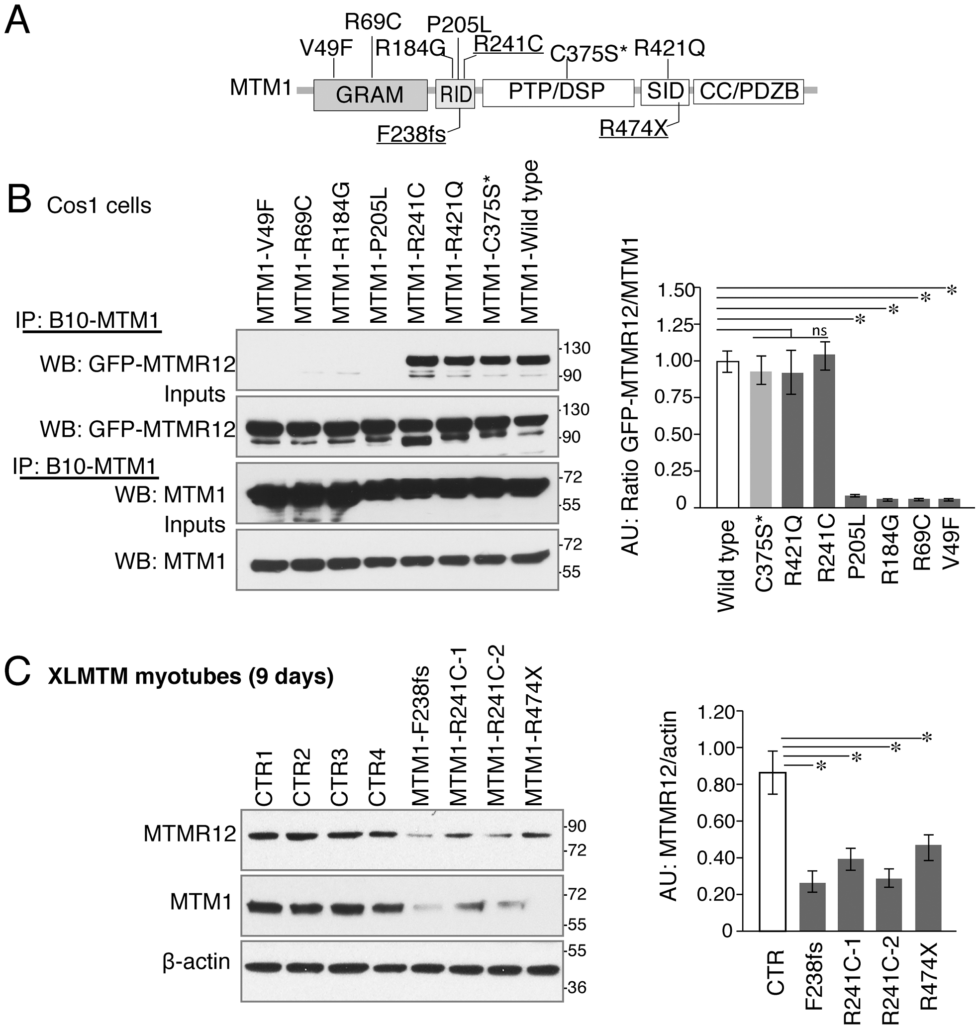
Myotubularin-MTMR12 interactions in XLMTM.
(A) Schematic diagram of different domains of myotubularin protein displaying representative pathogenic mutations found in XLMTM patients or an artificial inactivating mutation C375S* (GRAM, N terminal lipid or protein interacting domain; RID, putative membrane targeting motif; PTP/DSP, phosphatase domain; SID, protein-protein interacting domain; CC, coiled-coil domain; PDZB, PDZ binding site). (B) Wild-type or mutant MTM1-B10 fusion proteins with indicated missense mutations and wild-type MTMR12-GFP proteins were overexpressed in Cos1 cells. Immunoprecipitation of protein extracts with anti-B10 tag antibody showed that mutations on GRAM or RID domains disrupt the interactions between MTM1 and MTMR12. (C) Western blotting of XLMTM patient myotubes showed that mutants that decrease the stability of myotubularin protein also results in a reduction of MTMR12 levels. Histograms depict the western quantification for panels (B) and (C). Asterisks indicate statistically significant differences from measurements of wild type controls, Pd0.05.

