Fig. 7
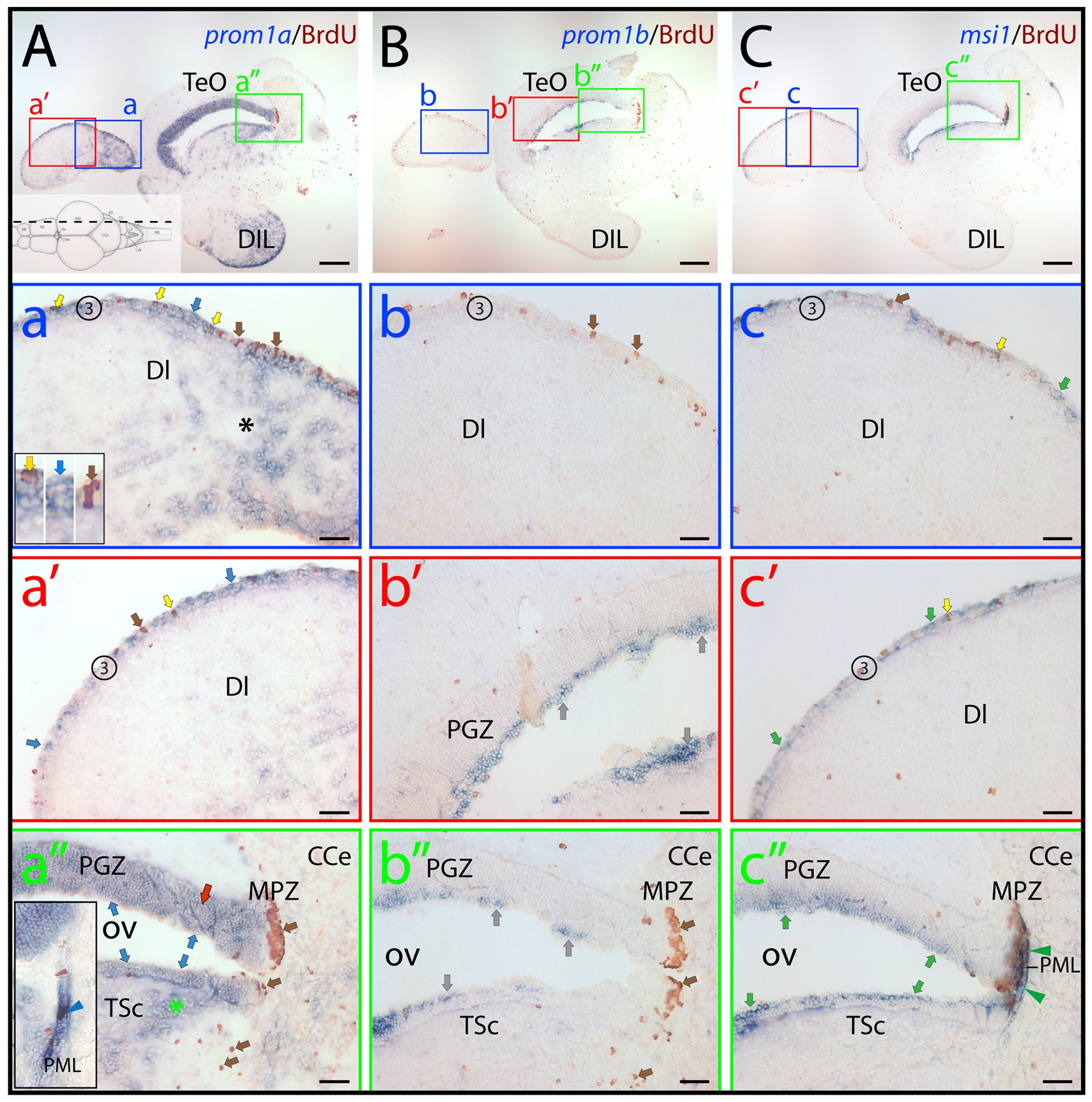
Distribution of zebrafish prominin-1a? and b?positive cells in the dorsal lateral telencephalon and the tectal ventricular zone.
(A?C) Cryosections of 3-month-old adult brain from BrdU-treated zebrafish were processed for ISH using an antisense DIG-labeled probe either against prominin-1a (A; prom1a), prominin-1b (B; prom1b) or musashi-1 (C; msi1). Proliferating cells were observed by immuno-detection of BrdU (brown). Position of paramedian longitudinal sections of the brain is indicated on the cartoon (A) adapted from the neuroanatomical atlas by Wulliman and colleagues [104]. The boxed areas in A, B and C are displayed at a higher magnification in panels a?a′′, b?b′′ and c?c′′, respectively. (A) Prominin-1a?positive cells are enriched in the diffuse nucleus of the hypothalamic inferior lobe (A, DIL) and in the extraventricular dorsal telencephalic parenchyma (a, black asterisk). They are found within the proliferative zone 3 (3) of the dorsal telencephalic surface (a, a′, blue arrows), and are either BrdU?positive or negative (yellow and blue arrows, respectively, see inset in a). Rare proliferating cells are devoid of prominin-1a (a, brown arrow). Prominin-1a?positive cells are also detected in the superficial (blue arrows) and deeper (red arrow and green asterisk) areas of periventricular grey zone (PGZ) and Torus semicircularis (TSc) lining the optic (tectal) ventricle (OV) (a′′), respectively. While the marginal proliferating zone of the tectum (MPZ) lacks prominin-1a, a small number of prominin-1a?positive proliferating cells is observed in the posterior mesencephalic lamina (PML) (a′′, see inset, blue arrowhead). (B) Prominin-1b?positive cells (grey arrows) are found in the superficial tear of PGZ (b′) and in TSc lining the OV (b′′), whereas proliferating cells within the zone 3 of the dorsal telencephalic surface (b, brown arrows), the deeper areas of the PGZ (b′) and in the MPZ (b′′) are negative. (C) Msi-1?positive cells found within the proliferative zone 3 of the dorsal telencephalic surface (c, c′) are either BrdU?positive or negative (yellow and green arrows, respectively). Rare proliferating cells are devoid of msi-1 (c, brown arrow). Msi-1 is detected in the superficial tear of PGZ and TSc lining the OV (c′′) as well as in proliferating cells of the MPZ and those of the PML (c′′, green arrowheads). CCe, corpus cerebelli; DI, dorsal lateral subdivision of the dorsal telencephalic region; TeO, tectum opticum. Scale bars, A?C, 250 μm; a?c′′, 50 μm.

