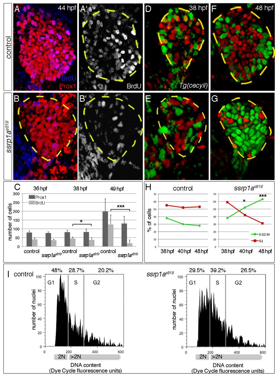
Fig. 3 Ssrp1a promotes cell cycle progression. (A-C) BrdU incorporation is reduced in ssrp1as819 mutant livers from 38 hpf onwards (outlined). Error bars represent s.d. (D-G) Transgenic cecyil expression marks G1 phase cells in red and S/G2/M phase cells in green. (H) Quantification of hepatoblast proliferation shows a reversed distribution in controls and ssrp1as819 mutants at 48 hpf. (I) FACS analysis of 42-44 hpf foregut endoderm shows an increase of cells in S phase and a decrease of those in G1 phase in ssrp1as819 mutants. A-B′,D-G are confocal projections of ventral views; all anterior to the top. *P<0.05, ***P<0.0005, determined by unpaired Student?s t-test.
Image
Figure Caption
Figure Data
Acknowledgments
This image is the copyrighted work of the attributed author or publisher, and
ZFIN has permission only to display this image to its users.
Additional permissions should be obtained from the applicable author or publisher of the image.
Full text @ Development

