
|
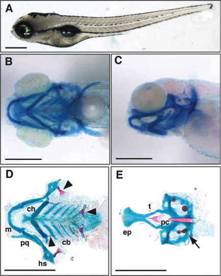
Fig. 1 External morphology and craniofacial cartilage organization in wild-type 5-day post-fertilization (dpf) Danio rerio larvae. A: Lateral view of a fixed zebrafish larva. B: Ventral view of larval head skeleton. C: Lateral view of same fish imaged in B. D: Flat-mount of viscerocranial skeleton after removal of the neurocranium (100� magnification). E: Flat-mount of neurocranium (100� magnification). cb, ceratobranchials; ch, ceratohyal; ep, ethmoid plate; hs, hyosymplectic; m, Meckel′s; pc, parachordals; pq, palatoquadrate; and t, trabecula. Bone and pharyngeal teeth (arrowheads) and otic vesicles (arrow) are also shown. Scale bars = 500 μm.