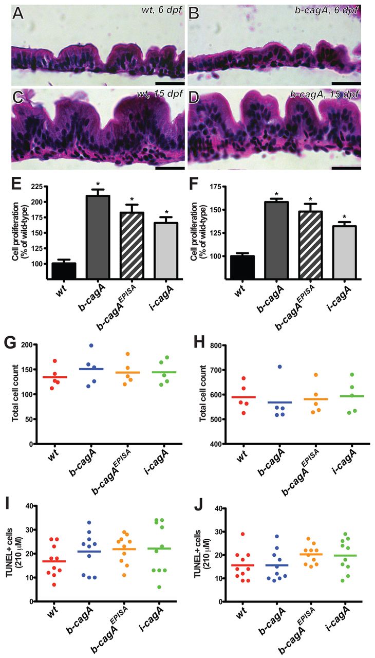
Fig. 2 CagA expression causes overproliferation of the intestinal epithelium. (A,B) H&E stained sagittal sections of wild-type (A) and b-cagA transgenic (B) zebrafish intestine at 6 dpf. (C,D) H&E stained sagittal sections of wild-type (C) and b-cagA transgenic (D) zebrafish intestine at 15 dpf. (E,F) Intestinal epithelial cell proliferation at 6 dpf (E) and 15 dpf (F). Bars represent proliferation (mean ± s.e.m.) as a percentage of wild-type; n=10, *P<0.05 using one-way ANOVA with Tukey?s test. (G,H) Total intestinal epithelial cell counts of single H&E stained midline sagittal sections at 6 dpf (G) and 15 dpf (H). (I,J) TUNEL-positive cells in the intestinal epithelium at 6 dpf (I) and 15 dpf (J). Scale bars: 10 μm.
Image
Figure Caption
Figure Data
Acknowledgments
This image is the copyrighted work of the attributed author or publisher, and
ZFIN has permission only to display this image to its users.
Additional permissions should be obtained from the applicable author or publisher of the image.
Full text @ Dis. Model. Mech.

