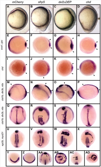
Fig. 2 Overexpression of sfrp5 disrupts gastrulation.
Embryos were injected with 100 pg mCherry as control (A, E, I, M, Q, U, Y, AA, AC), 140 pg sfrp5 (B, F, J, N, R, V, Z, AB, AD), 150 pg dvl2?DEP (C, G, K, O, S, W), or 50 pg chd (D, H, L, P, T, X). A?D) Morphology of injected embryos injected at early somitogenesis, lateral view with dorsal side to right. E?AD) Whole-mount in situ hybridization of injected embryos. E?H) Animal pole view with dorsal side to the right of embryos stained with eve1 and gsc probes at shield stage. Arrowheads demarcate gsc staining. I?L) Animal pole view with dorsal side to the right of embryos stained with chd, demarcated by arrowheads, at shield stage. M?T) Early somitogenesis embryos stained with probes against ctsl1b, dlx3b, and ntla. M?P: Lateral view with dorsal to top. Arrowheads mark length of notochord. Q?T: dorsal view with anterior to top. Brackets show notochord width. U?X) Mid-somitogenesis embryos stained with egr2b and myoD1. Dorsal view with anterior to top. In X, arrows mark radialization of egfr2b and myoD1 staining. Y, Z) Bud-stage embryos hybridized with probe against her5; anterior view, dorsal to bottom. AA?AB) Early somitogenesis embryos hybridized with probes against ctsl1b, dlx3b, and ntla. Arrow: normal ctsl1b staining. Arrowhead: ectopic ctsl1b staining. AA: Dorsal view, anterior to top. AB: Ventral view, dorsal to top. AC?AD) Mid-somitogenesis embryos hybridized with probes against egr2b and myoD1. AC: Dorsal view, anterior to top. AD: ventral view, dorsal to top. Arrows point to rhombomere 3.

