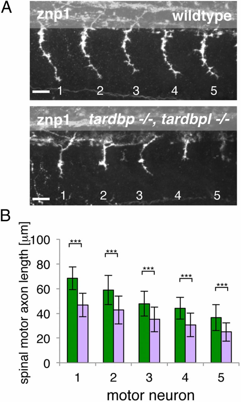
Fig. 5 Spinal motor neuron axon outgrowth phenotype of tardbp-/-;tardbpl-/- mutants. (A) Znp-1 antibody staining of motor neuron axons in 28-hpf-old embryos of the five somites (labeled 1?5) anterior to the end of the yolk extension. Spinal motor neuron axons of tardbp-/-;tardbpl-/- mutants are reduced in length compared with wild-type siblings. Anterior to the left, lateral view. (Scale bar, 25 μm.) (B) Quantitation of the length of the outgrowing spinal motor neuron axons measured from the exit point of the spinal cord to the growth cone in wild-type (green) and tardbp-/-;tardbpl-/- mutants (purple). Error bars indicate ąSD, n ≥ 13 embryos per experiment, ***P < 0.0015, Student t test.
Image
Figure Caption
Acknowledgments
This image is the copyrighted work of the attributed author or publisher, and
ZFIN has permission only to display this image to its users.
Additional permissions should be obtained from the applicable author or publisher of the image.
Full text @ Proc. Natl. Acad. Sci. USA

