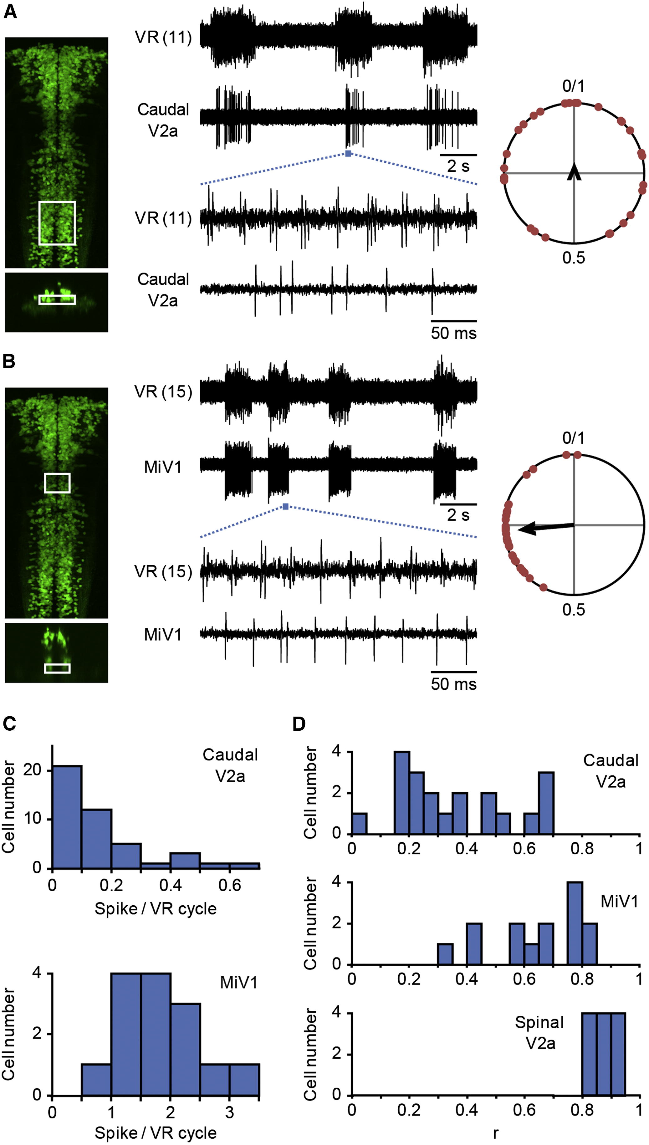
Fig. 5 Activity of Hindbrain V2a Neurons during Fictive Swimming Compound transgenic fish of Tg[chx10:Gal4] and Tg[UAS:GFP] at 3 dpf were used for electrophysiological recordings. Loose-patch recordings from reticulospinal V2a neurons were made together with VR recordings. (A and B) (A) shows an example of recordings from the small reticulospinal V2a neurons in the caudal hindbrain, whereas (B) shows an example of recordings from the MiV1 neurons. The left panels show the region of the cells that were recorded. The top panel for each figure is a dorsal view, whereas the bottom panel is a cross-section. Middle panels show recordings. The numbers in parentheses indicate the locations of the VR recordings. The right panels show circular plots that depict the phasic relation of V2a neuron spikes to the VR activity. The central time point of a VR activity was assigned a phase value of zero and that of the next VR activity was assigned a phase value of one (Figure S5 C). The phase values of 30 randomly selected spikes are plotted in the circle. The direction of the vector shows the mean of the phase value, whereas the length of the vector shows the strength of the rhythmicity. (C) Histograms of the number of spikes of reticulospinal V2a neurons per VR cycle. The top panel shows a histogram of the small reticulospinal V2a neurons in the caudal hindbrain, whereas the bottom panel shows a histogram of the MiV1 neurons. n = 44 for the former, and n = 14 for the latter. (D) Histograms of the length of the vectors (“r”). The top panel shows the histogram of the small reticulospinal V2a neurons in the caudal hindbrain. In this histogram, only those neurons whose firing frequencies were more than 0.1 are included. n = 20. The middle panel shows the histogram of the MiV1 neurons. n = 14. The bottom panel shows the histogram of the spinal V2a neurons. This figure is shown for comparison purposes. n = 12.
Image
Figure Caption
Acknowledgments
This image is the copyrighted work of the attributed author or publisher, and
ZFIN has permission only to display this image to its users.
Additional permissions should be obtained from the applicable author or publisher of the image.
Full text @ Curr. Biol.

